
The radiological evolution after COVID-19 pneumonia is unknown. We propose to analyze the main radiological findings one year after COVID-19 pneumonia, as well as identify possible factors that may influence it.
Material and methodsCohort study of patients with COVID-19 pneumonia undergoing high-resolution computed tomography (HRCT) 12 months after hospital discharge. A descriptive study of the radiological findings and a multivariate analysis are carried out to identify the factors of the appearance of radiological anomalies.
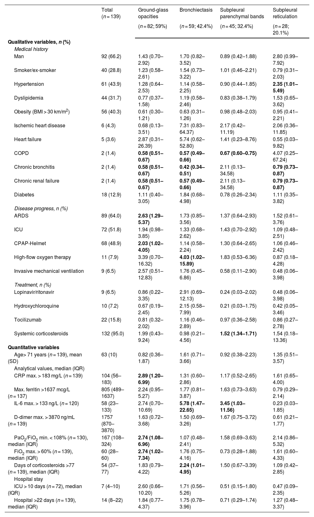
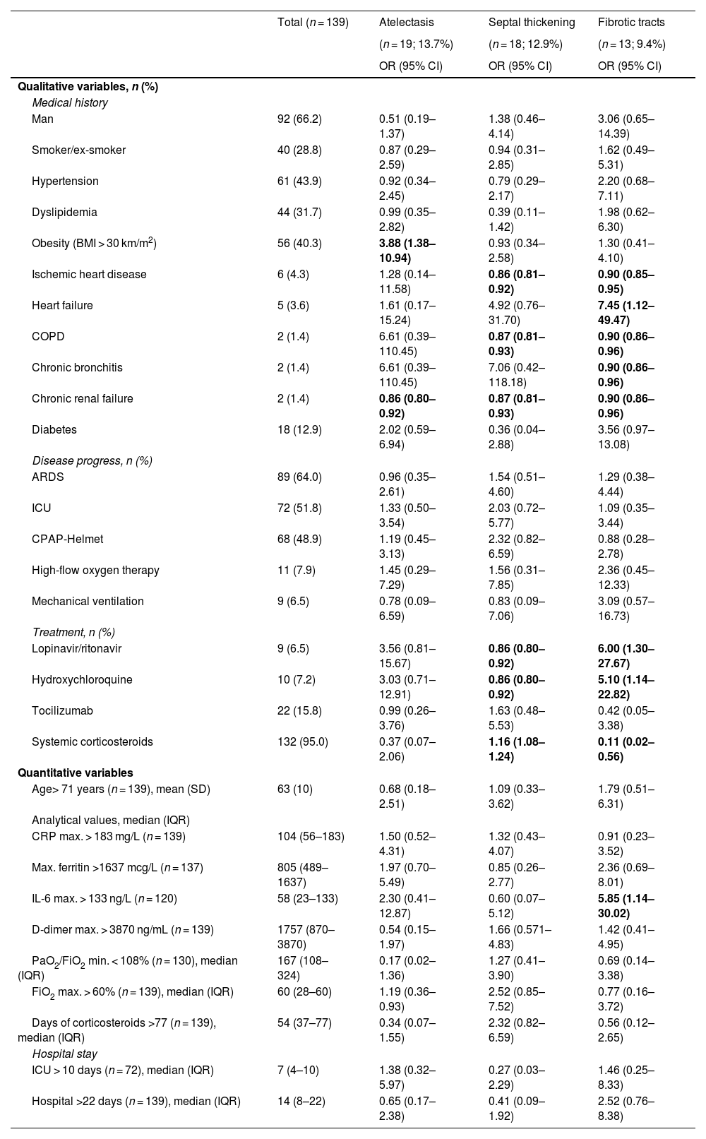
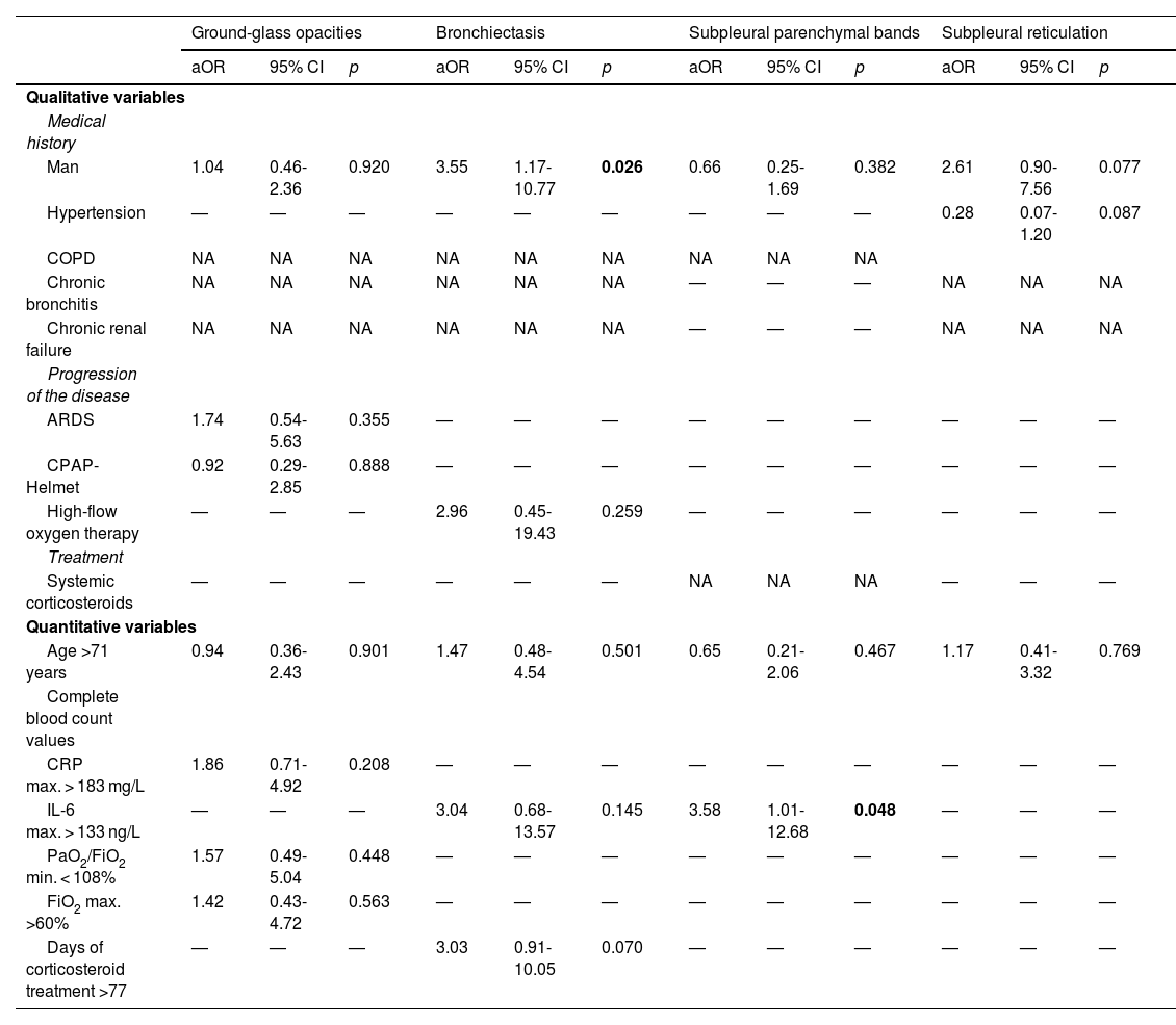
Resultsn = 139 patients, with a mean age of 63 years and 66.2% male. The most frequent radiological findings were ground-glass opacities (59%), followed by bronchiectasis (42.4%), subpleural parenchymal bands (32.4%), atelectasis (13.7%), septal thickening (12.9%) and fibrotic tracts (9.4%). Male sex was associated with the presence of bronchiectasis (ORa = 3.55; p = 0.026), peak admission IL-6 levels > 133 ng/L with the detection of subpleural parenchymal bands (ORa = 3.58; p = 0.048) and obesity with the occurrence of atelectasis (ORa = 3.70; p = 0.014). Systemic corticotherapy during admission decreased the risk of fibrotic tracts (ORa = 0.02; p = 0.003).
ConclusionsLung damage persists with high frequency one year after COVID-19 pneumonia. Male sex, high peak admission IL-6 levels and obesity were risk factors for radiological abnormalities while systemic corticosteroid therapy decreased the occurrence of fibrotic tracts 12 months after hospital admission.
La evolución radiológica tras una neumonía COVID-19 es desconocida. Nos proponemos analizar cuáles son los principales hallazgos radiológicos al año de una neumonía COVID-19, así como identificar posibles factores que puedan influir en ello.
Materiales y métodosEstudio de cohortes de pacientes con neumonía por COVID-19 a los que se les hace una tomografía computarizada de alta resolución (TACAR) a los 12 meses del alta hospitalaria. Se realiza estudio descriptivo de los hallazgos radiológicos y un análisis multivariante para identificar los factores de la aparición de anomalías radiológicas.
Resultadosn = 139 pacientes, con una media de edad de 63 años y 66,2% varones. Los hallazgos radiológicos más frecuentes fueron las opacidades en vidrio deslustrado (59%), seguido de bronquiectasias (42,4%), bandas parenquimatosas subpleurales (32,4%), atelectasias (13,7%), engrosamiento septal (12,9%) y tractos fibróticos (9,4%). El sexo masculino se asoció a la presencia de bronquiectasias (ORa = 3,55; p = 0,026), los niveles máximos de IL-6 durante el ingreso > 133 ng/L a la detección de bandas parenquimatosas subpleurales (ORa = 3,58; p = 0,048) y la obesidad con la aparición de atelectasias (ORa = 3,70; p = 0,014). La corticoterapia sistémica durante el ingreso disminuyó el riesgo de aparición de tractos fibróticos (ORa = 0,02; p = 0,003).
Conclusionesel daño pulmonar persiste con elevada frecuencia un año después de la neumonía COVID-19. El sexo masculino, los niveles máximos de IL-6 durante el ingreso elevados y la obesidad se comportaron como factores de riesgo para presentar anomalías radiológicas mientras que la corticoterapia sistémica disminuyó la aparición de tractos fibróticos a los 12 meses del ingreso hospitalario.
Article
Diríjase desde aquí a la web de la >>>FESEMI<<< e inicie sesión mediante el formulario que se encuentra en la barra superior, pulsando sobre el candado.

Una vez autentificado, en la misma web de FESEMI, en el menú superior, elija la opción deseada.
>>>FESEMI<<<


