Share
array:24 [
"pii" => "S225488742100031X"
"issn" => "22548874"
"doi" => "10.1016/j.rceng.2020.01.006"
"estado" => "S300"
"fechaPublicacion" => "2021-05-01"
"aid" => "1871"
"copyright" => "Elsevier España, S.L.U. and Sociedad Española de Medicina Interna (SEMI)"
"copyrightAnyo" => "2020"
"documento" => "article"
"crossmark" => 1
"subdocumento" => "fla"
"cita" => "Rev Clin Esp. 2021;221:258-63"
"abierto" => array:3 [
"ES" => false
"ES2" => false
"LATM" => false
]
"gratuito" => false
"lecturas" => array:1 [
"total" => 0
]
"Traduccion" => array:1 [
"es" => array:19 [
"pii" => "S0014256520301521"
"issn" => "00142565"
"doi" => "10.1016/j.rce.2020.01.012"
"estado" => "S300"
"fechaPublicacion" => "2021-05-01"
"aid" => "1871"
"copyright" => "Elsevier España, S.L.U. and Sociedad Española de Medicina Interna (SEMI)"
"documento" => "article"
"crossmark" => 1
"subdocumento" => "fla"
"cita" => "Rev Clin Esp. 2021;221:258-63"
"abierto" => array:3 [
"ES" => false
"ES2" => false
"LATM" => false
]
"gratuito" => false
"lecturas" => array:1 [
"total" => 0
]
"es" => array:12 [
"idiomaDefecto" => true
"cabecera" => "<span class="elsevierStyleTextfn">ORIGINAL</span>"
"titulo" => "Validez de la ecografía torácica para descartar neumotórax iatrogénico realizada por neumólogos sin experiencia en este procedimiento"
"tienePdf" => "es"
"tieneTextoCompleto" => "es"
"tieneResumen" => array:2 [
0 => "es"
1 => "en"
]
"paginas" => array:1 [
0 => array:2 [
"paginaInicial" => "258"
"paginaFinal" => "263"
]
]
"titulosAlternativos" => array:1 [
"en" => array:1 [
"titulo" => "Validity of lung ultrasound to rule out iatrogenic pneumothorax performed by pulmonologists without experience in this procedure"
]
]
"contieneResumen" => array:2 [
"es" => true
"en" => true
]
"contieneTextoCompleto" => array:1 [
"es" => true
]
"contienePdf" => array:1 [
"es" => true
]
"autores" => array:1 [
0 => array:2 [
"autoresLista" => "C. Ramos Hernández, M. Núñez Delgado, M. Botana Rial, C. Mouronte Roibás, V. Leiro Fernández, C. Vilariño Pombo, R. Tubío Pérez, M. Nuñez Fernández, A. Fernández Villar"
"autores" => array:9 [
0 => array:2 [
"nombre" => "C."
"apellidos" => "Ramos Hernández"
]
1 => array:2 [
"nombre" => "M."
"apellidos" => "Núñez Delgado"
]
2 => array:2 [
"nombre" => "M."
"apellidos" => "Botana Rial"
]
3 => array:2 [
"nombre" => "C."
"apellidos" => "Mouronte Roibás"
]
4 => array:2 [
"nombre" => "V."
"apellidos" => "Leiro Fernández"
]
5 => array:2 [
"nombre" => "C."
"apellidos" => "Vilariño Pombo"
]
6 => array:2 [
"nombre" => "R."
"apellidos" => "Tubío Pérez"
]
7 => array:2 [
"nombre" => "M."
"apellidos" => "Nuñez Fernández"
]
8 => array:2 [
"nombre" => "A."
"apellidos" => "Fernández Villar"
]
]
]
]
]
"idiomaDefecto" => "es"
"Traduccion" => array:1 [
"en" => array:9 [
"pii" => "S225488742100031X"
"doi" => "10.1016/j.rceng.2020.01.006"
"estado" => "S300"
"subdocumento" => ""
"abierto" => array:3 [
"ES" => false
"ES2" => false
"LATM" => false
]
"gratuito" => false
"lecturas" => array:1 [
"total" => 0
]
"idiomaDefecto" => "en"
"EPUB" => "https://multimedia.elsevier.es/PublicationsMultimediaV1/item/epub/S225488742100031X?idApp=WRCEE"
]
]
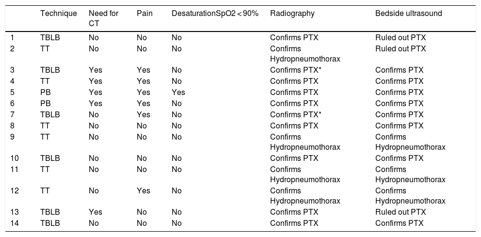
"EPUB" => "https://multimedia.elsevier.es/PublicationsMultimediaV1/item/epub/S0014256520301521?idApp=WRCEE"
"url" => "/00142565/0000022100000005/v1_202105061437/S0014256520301521/v1_202105061437/es/main.assets"
]
]
"itemSiguiente" => array:19 [
"pii" => "S2254887421000333"
"issn" => "22548874"
"doi" => "10.1016/j.rceng.2020.10.005"
"estado" => "S300"
"fechaPublicacion" => "2021-05-01"
"aid" => "1918"
"copyright" => "Elsevier España, S.L.U. and Sociedad Española de Medicina Interna (SEMI)"
"documento" => "article"
"crossmark" => 1
"subdocumento" => "fla"
"cita" => "Rev Clin Esp. 2021;221:264-73"
"abierto" => array:3 [
"ES" => false
"ES2" => false
"LATM" => false
]
"gratuito" => false
"lecturas" => array:1 [
"total" => 0
]
"en" => array:13 [
"idiomaDefecto" => true
"cabecera" => "<span class="elsevierStyleTextfn">Original article</span>"
"titulo" => "Differential risk for COVID-19 in the first wave of the disease among Spaniards and migrants from different areas of the world living in Spain"
"tienePdf" => "en"
"tieneTextoCompleto" => "en"
"tieneResumen" => array:2 [
0 => "en"
1 => "es"
]
"paginas" => array:1 [
0 => array:2 [
"paginaInicial" => "264"
"paginaFinal" => "273"
]
]
"titulosAlternativos" => array:1 [
"es" => array:1 [
"titulo" => "Riesgo de COVID-19 en españoles y migrantes de distintas zonas del mundo residentes en España en la primera oleada de la enfermedad"
]
]
"contieneResumen" => array:2 [
"en" => true
"es" => true
]
"contieneTextoCompleto" => array:1 [
"en" => true
]
"contienePdf" => array:1 [
"en" => true
]
"resumenGrafico" => array:2 [
"original" => 0
"multimedia" => array:8 [
"identificador" => "fig0010"
"etiqueta" => "Figure 2"
"tipo" => "MULTIMEDIAFIGURA"
"mostrarFloat" => true
"mostrarDisplay" => false
"figura" => array:1 [
0 => array:4 [
"imagen" => "gr2.jpeg"
"Alto" => 2710
"Ancho" => 2508
"Tamanyo" => 275327
]
]
"detalles" => array:1 [
0 => array:3 [
"identificador" => "at0010"
"detalle" => "Figure "
"rol" => "short"
]
]
"descripcion" => array:1 [
"en" => "<p id="spar0020" class="elsevierStyleSimplePara elsevierViewall">Relative risks for COVID-19, calculated by means of a negative binomial regression analysis.</p> <p id="spar0025" class="elsevierStyleSimplePara elsevierViewall"><span class="elsevierStyleItalic">Panel A:</span> relative risks for COVID-19 according to age intervals (reference age < 30 years) and sex.</p> <p id="spar0030" class="elsevierStyleSimplePara elsevierViewall"><span class="elsevierStyleItalic">Panel B:</span> world area of origin (reference area: Spain).</p>"
]
]
]
"autores" => array:1 [
0 => array:2 [
"autoresLista" => "C. Guijarro, E. Pérez-Fernández, B. González-Piñeiro, V. Meléndez, M.J. Goyanes, M.E. Renilla, M.L. Casas, I. Sastre, M. Velasco"
"autores" => array:10 [
0 => array:2 [
"nombre" => "C."
"apellidos" => "Guijarro"
]
1 => array:2 [
"nombre" => "E."
"apellidos" => "Pérez-Fernández"
]
2 => array:2 [
"nombre" => "B."
"apellidos" => "González-Piñeiro"
]
3 => array:2 [
"nombre" => "V."
"apellidos" => "Meléndez"
]
4 => array:2 [
"nombre" => "M.J."
"apellidos" => "Goyanes"
]
5 => array:2 [
"nombre" => "M.E."
"apellidos" => "Renilla"
]
6 => array:2 [
"nombre" => "M.L."
"apellidos" => "Casas"
]
7 => array:2 [
"nombre" => "I."
"apellidos" => "Sastre"
]
8 => array:2 [
"nombre" => "M."
"apellidos" => "Velasco"
]
9 => array:1 [
"colaborador" => "the Alcorcón COVID Investigators"
]
]
]
]
]
"idiomaDefecto" => "en"
"Traduccion" => array:1 [
"es" => array:9 [
"pii" => "S0014256520302976"
"doi" => "10.1016/j.rce.2020.10.006"
"estado" => "S300"
"subdocumento" => ""
"abierto" => array:3 [
"ES" => false
"ES2" => false
"LATM" => false
]
"gratuito" => false
"lecturas" => array:1 [
"total" => 0
]
"idiomaDefecto" => "es"
"EPUB" => "https://multimedia.elsevier.es/PublicationsMultimediaV1/item/epub/S0014256520302976?idApp=WRCEE"
]
]
"EPUB" => "https://multimedia.elsevier.es/PublicationsMultimediaV1/item/epub/S2254887421000333?idApp=WRCEE"
"url" => "/22548874/0000022100000005/v3_202105110748/S2254887421000333/v3_202105110748/en/main.assets"
]
"itemAnterior" => array:19 [
"pii" => "S2254887421000382"
"issn" => "22548874"
"doi" => "10.1016/j.rceng.2020.01.009"
"estado" => "S300"
"fechaPublicacion" => "2021-05-01"
"aid" => "1864"
"copyright" => "Elsevier España, S.L.U. and Sociedad Española de Medicina Interna (SEMI)"
"documento" => "article"
"crossmark" => 1
"subdocumento" => "fla"
"cita" => "Rev Clin Esp. 2021;221:249-57"
"abierto" => array:3 [
"ES" => false
"ES2" => false
"LATM" => false
]
"gratuito" => false
"lecturas" => array:1 [
"total" => 0
]
"en" => array:13 [
"idiomaDefecto" => true
"cabecera" => "<span class="elsevierStyleTextfn">Original article</span>"
"titulo" => "Risk factors and comorbidities associated with severe aortic stenosis: A case-control study"
"tienePdf" => "en"
"tieneTextoCompleto" => "en"
"tieneResumen" => array:2 [
0 => "en"
1 => "es"
]
"paginas" => array:1 [
0 => array:2 [
"paginaInicial" => "249"
"paginaFinal" => "257"
]
]
"titulosAlternativos" => array:1 [
"es" => array:1 [
"titulo" => "Factores de riesgo y comorbilidades asociadas a la estenosis aórtica grave: estudio de casos y controles"
]
]
"contieneResumen" => array:2 [
"en" => true
"es" => true
]
"contieneTextoCompleto" => array:1 [
"en" => true
]
"contienePdf" => array:1 [
"en" => true
]
"resumenGrafico" => array:2 [
"original" => 0
"multimedia" => array:8 [
"identificador" => "fig0010"
"etiqueta" => "Figure 2"
"tipo" => "MULTIMEDIAFIGURA"
"mostrarFloat" => true
"mostrarDisplay" => false
"figura" => array:1 [
0 => array:4 [
"imagen" => "gr2.jpeg"
"Alto" => 1504
"Ancho" => 1483
"Tamanyo" => 71776
]
]
"detalles" => array:1 [
0 => array:3 [
"identificador" => "at0010"
"detalle" => "Figure "
"rol" => "short"
]
]
"descripcion" => array:1 [
"en" => "<p id="spar0010" class="elsevierStyleSimplePara elsevierViewall">ROC curve of the multivariate logistic regression model for comorbidities. ROC = 0.74 (0.68–0.80).</p>"
]
]
]
"autores" => array:1 [
0 => array:2 [
"autoresLista" => "J.M. Gracia Baena, I. Calaf Vall, M. Zielonka, J.R. Marsal Mora, P. Godoy, F. Worner Diz"
"autores" => array:6 [
0 => array:2 [
"nombre" => "J.M."
"apellidos" => "Gracia Baena"
]
1 => array:2 [
"nombre" => "I."
"apellidos" => "Calaf Vall"
]
2 => array:2 [
"nombre" => "M."
"apellidos" => "Zielonka"
]
3 => array:2 [
"nombre" => "J.R."
"apellidos" => "Marsal Mora"
]
4 => array:2 [
"nombre" => "P."
"apellidos" => "Godoy"
]
5 => array:2 [
"nombre" => "F."
"apellidos" => "Worner Diz"
]
]
]
]
]
"idiomaDefecto" => "en"
"Traduccion" => array:1 [
"es" => array:9 [
"pii" => "S0014256520301454"
"doi" => "10.1016/j.rce.2020.01.010"
"estado" => "S300"
"subdocumento" => ""
"abierto" => array:3 [
"ES" => false
"ES2" => false
"LATM" => false
]
"gratuito" => false
"lecturas" => array:1 [
"total" => 0
]
"idiomaDefecto" => "es"
"EPUB" => "https://multimedia.elsevier.es/PublicationsMultimediaV1/item/epub/S0014256520301454?idApp=WRCEE"
]
]
"EPUB" => "https://multimedia.elsevier.es/PublicationsMultimediaV1/item/epub/S2254887421000382?idApp=WRCEE"
"url" => "/22548874/0000022100000005/v3_202105110748/S2254887421000382/v3_202105110748/en/main.assets"
]
"en" => array:19 [
"idiomaDefecto" => true
"cabecera" => "<span class="elsevierStyleTextfn">Original article</span>"
"titulo" => "Validity of lung ultrasound to rule out iatrogenic pneumothorax performed by pulmonologists without experience in this procedure"
"tieneTextoCompleto" => true
"paginas" => array:1 [
0 => array:2 [
"paginaInicial" => "258"
"paginaFinal" => "263"
]
]
"autores" => array:1 [
0 => array:4 [
"autoresLista" => "C. Ramos Hernández, M. Núñez Delgado, M. Botana Rial, C. Mouronte Roibás, V. Leiro Fernández, C. Vilariño Pombo, R. Tubío Pérez, M. Nuñez Fernández, A. Fernández Villar"
"autores" => array:9 [
0 => array:4 [
"nombre" => "C."
"apellidos" => "Ramos Hernández"
"email" => array:1 [
0 => "cristina.ramos.hernandez@sergas.es"
]
"referencia" => array:1 [
0 => array:2 [
"etiqueta" => "*"
"identificador" => "cor0005"
]
]
]
1 => array:2 [
"nombre" => "M."
"apellidos" => "Núñez Delgado"
]
2 => array:2 [
"nombre" => "M."
"apellidos" => "Botana Rial"
]
3 => array:2 [
"nombre" => "C."
"apellidos" => "Mouronte Roibás"
]
4 => array:2 [
"nombre" => "V."
"apellidos" => "Leiro Fernández"
]
5 => array:2 [
"nombre" => "C."
"apellidos" => "Vilariño Pombo"
]
6 => array:2 [
"nombre" => "R."
"apellidos" => "Tubío Pérez"
]
7 => array:2 [
"nombre" => "M."
"apellidos" => "Nuñez Fernández"
]
8 => array:2 [
"nombre" => "A."
"apellidos" => "Fernández Villar"
]
]
"afiliaciones" => array:1 [
0 => array:2 [
"entidad" => "Servicio de Neumología. Hospital Álvaro Cunqueiro. EOXI de Vigo PneumoVigo I+i. Instituto de Investigación Sanitaria Galicia Sur (IISGS), Vigo, Spain"
"identificador" => "aff0005"
]
]
"correspondencia" => array:1 [
0 => array:3 [
"identificador" => "cor0005"
"etiqueta" => "⁎"
"correspondencia" => "Corresponding author."
]
]
]
]
"titulosAlternativos" => array:1 [
"es" => array:1 [
"titulo" => "Validez de la ecografía torácica para descartar neumotórax iatrogénico realizada por neumólogos sin experiencia en este procedimiento"
]
]
"textoCompleto" => "<span class="elsevierStyleSections"><span id="sec0005" class="elsevierStyleSection elsevierViewall"><span class="elsevierStyleSectionTitle" id="sect0065">Introduction</span><p id="par0005" class="elsevierStylePara elsevierViewall">Lung ultrasound (LUS) has been shown to have clear benefits for respiratory pathology studies compared with other imaging techniques, including speed, as it enables bedside examination, and is low cost, emits no radiation, and offers the ability to produce moving images. This has represented a shift in clinical practice and the number of applications is increasingly expanding.<a class="elsevierStyleCrossRef" href="#bib0005"><span class="elsevierStyleSup">1</span></a></p><p id="par0010" class="elsevierStylePara elsevierViewall">Ultrasound is a useful technique for evaluating pleural effusion, identifying peripheral lung lesions, pneumonia or interstitial alterations.<a class="elsevierStyleCrossRef" href="#bib0010"><span class="elsevierStyleSup">2</span></a> LUS is also a useful tool for diagnosing pneumothorax and can even be more sensitive than chest radiography when performed by expert radiologists. The published meta-analyses show a sensitivity of about 80% versus 40-50% when compared with radiography, though the specificity is similar between the two techniques, this being 99%.<a class="elsevierStyleCrossRefs" href="#bib0015"><span class="elsevierStyleSup">3,4</span></a></p><p id="par0015" class="elsevierStylePara elsevierViewall">Therefore, implementation of ultrasonography is progressive and increasingly more specialists are regularly incorporating its use. In the United Kingdom, knowledge of imaging techniques, including LUS, is included in training for pulmonologists.<a class="elsevierStyleCrossRef" href="#bib0025"><span class="elsevierStyleSup">5</span></a></p><p id="par0020" class="elsevierStylePara elsevierViewall">The current recommendations in manuals and guidelines are to perform chest radiography following semi-invasive bronchopleural procedures to rule out complications such as pneumothorax, particularly in symptomatic patients.<a class="elsevierStyleCrossRefs" href="#bib0030"><span class="elsevierStyleSup">6–8</span></a> LUS conducted by the pulmonologist who performed the initial procedure could be of use for early detection of pneumothorax, preventing unnecessary chest radiography and its resulting radiation exposure. However, few studies have analysed this aspect, particularly when the ultrasound is not performed by experts with ultrasound training.<a class="elsevierStyleCrossRefs" href="#bib0045"><span class="elsevierStyleSup">9–11</span></a></p><p id="par0025" class="elsevierStylePara elsevierViewall">The main aim of this study was therefore to determine the validity of LUS in ruling out pneumothorax after bronchopleural procedures conducted by pulmonologists without specific LUS training to that end and without the support of radiologists.</p></span><span id="sec0010" class="elsevierStyleSection elsevierViewall"><span class="elsevierStyleSectionTitle" id="sect0070">Material and methods</span><p id="par0030" class="elsevierStylePara elsevierViewall">Prospective, observational study performed in consecutive patients from December 2014 to December 2015. An average of 450 pleural procedures and 1,550 endoscopic techniques are performed per year. We included in the study all patients who underwent a transbronchial lung biopsy (TBLB) without fluoroscopic guidance, therapeutic thoracentesis (TT) or transparietal pleural biopsy (PB) for whom, according to our unit guidelines, post-procedure radiography would be indicated.</p><p id="par0035" class="elsevierStylePara elsevierViewall">Exclusion criteria included being under 18 years of age, pregnant women, lack of informed consent, if any of the five participating pulmonologists were not present, and cases with severe complications that required emergency treatment procedures during or immediately after the intervention.</p><p id="par0040" class="elsevierStylePara elsevierViewall">The TBLBs were performed using a flexible bronchoscope (IT20; Olympus) under conscious sedation and local anaesthesia with the patient in the supine position. Samples were taken using oval forceps and between four and six biopsies were taken in the most affected area shown in the computerised tomography (CT).<a class="elsevierStyleCrossRef" href="#bib0050"><span class="elsevierStyleSup">10</span></a></p><p id="par0045" class="elsevierStylePara elsevierViewall">The TTs and PBs were performed with the patient in a sitting position and using topical anaesthesia with 2% mepivacaine. One ultrasound machine was used to identify the best area for the puncture, using in this case a convex 2−5 MHz (Sonosite C60n) probe. The needles used to perform the PBs were an Abrams needle, Tru-cut needle or Cope needle depending on the CT findings and at the discretion of the pulmonologist in charge of the technique.<a class="elsevierStyleCrossRefs" href="#bib0060"><span class="elsevierStyleSup">12,13</span></a></p><p id="par0050" class="elsevierStylePara elsevierViewall">A 14 G fenestrated catheter was used for the TTs and an aspiration system was subsequently connected.</p><p id="par0055" class="elsevierStylePara elsevierViewall">The pleural and bronchial procedures were performed by five pulmonologists with more than eight years of specific experience dedicated to bronchopleural techniques as well as three 2nd to 4th year pulmonology residents under supervision. All were experienced in LUS for managing pleural effusion and, prior to their participation in the study, had received basic information about detecting pneumothorax using this technique. This training included a one-hour theoretical talk and assessment of at least three supervised ultrasounds. Records were not taken regarding which participating pulmonologist performed each procedure.</p><span id="sec0015" class="elsevierStyleSection elsevierViewall"><span class="elsevierStyleSectionTitle" id="sect0075">Lung ultrasound</span><p id="par0060" class="elsevierStylePara elsevierViewall">Immediately following the bronchopleural technique, the same physician who conducted the test performed LUS.</p><p id="par0065" class="elsevierStylePara elsevierViewall">The patient was examined in a supine position with the head elevated to 45°, visualising the 2nd and 3rd intercostal space of each hemithorax using a portable ultrasound machine (Sonosite Fujifilm M-turbo) and a linear 5−10 MHz (Sonosite L38XI) transducer.</p><p id="par0070" class="elsevierStylePara elsevierViewall">The presence or absence of these signs were recorded in the following order in a case report form specifically designed for the study: lung sliding → B-lines → barcode → lung point.</p><p id="par0075" class="elsevierStylePara elsevierViewall">Pneumothorax was considered confirmed via ultrasound in the presence of the "lung point" sign. In the absence of the lung point, we also considered pneumothorax to be confirmed via ultrasound in the presence of a combination of the following signs:<a class="elsevierStyleCrossRef" href="#bib0005"><span class="elsevierStyleSup">1</span></a><ul class="elsevierStyleList" id="lis0005"><li class="elsevierStyleListItem" id="lsti0005"><span class="elsevierStyleLabel">•</span><p id="par0080" class="elsevierStylePara elsevierViewall">Absence of lung sliding during respiration.</p></li><li class="elsevierStyleListItem" id="lsti0010"><span class="elsevierStyleLabel">•</span><p id="par0085" class="elsevierStylePara elsevierViewall">Absence of B-lines arising from the pleural line and reaching the lower edge of the screen, erasing the A-lines in their wake.</p></li></ul></p><p id="par0090" class="elsevierStylePara elsevierViewall">Barcode: ultrasound sign visualised in M mode taking the form of parallel, horizontal lines.</p><p id="par0095" class="elsevierStylePara elsevierViewall">Pneumothorax was suspected when only one of these signs was present in an isolated manner. If any of the signs were ambiguous, the ultrasound was deemed consistent with suspected pneumothorax. If none of the criteria was met, we deemed the ultrasound to have ruled out pneumothorax.</p><p id="par0100" class="elsevierStylePara elsevierViewall">The “gold standard” was defined as the findings shown on the postero-anterior chest radiography during inhalation described by an expert radiologist in chest radiology and performed in the two hours following the procedure or later if the radiology scan was repeated due to the presence of symptoms suggestive of this complication, as recommended in the clinical practice guidelines.<a class="elsevierStyleCrossRefs" href="#bib0035"><span class="elsevierStyleSup">7,8</span></a></p><p id="par0105" class="elsevierStylePara elsevierViewall">Other variables were collected including demographic data (age, sex), presence of obesity and history of prior chronic respiratory disease. Radiological alterations that could affect ultrasound interpretation, such as the presence of atelectasis, adhesions, or emphysema, were also recorded.</p><p id="par0110" class="elsevierStylePara elsevierViewall">The presence of symptoms following the bronchopleural procedure such as pain or dyspnoea were also noted.</p><p id="par0115" class="elsevierStylePara elsevierViewall">Informed consent was obtained from all the patients participating in the study to perform the bronchopleural techniques and authorisation was granted for data collection.</p><p id="par0120" class="elsevierStylePara elsevierViewall">The study was approved by the Pontevedra-Vigo-Ourense Research Ethics Committee, Registration Code: 2018/526.</p></span><span id="sec0020" class="elsevierStyleSection elsevierViewall"><span class="elsevierStyleSectionTitle" id="sect0080">Statistical analysis</span><p id="par0125" class="elsevierStylePara elsevierViewall">A descriptive analysis of the demographic variables and the isolated and grouped pneumothorax ultrasound signs was performed. Validity parameters for the diagnostic tests were calculated using contingency tables and standard formulas for each of the pneumothorax ultrasound signs. The overall results were expressed in absolute frequencies and percentages for the qualitative variables and as means plus standard deviation (SD) for the numerical variables. The comparison between variables was conducted using the <span class="elsevierStyleItalic">X</span><span class="elsevierStyleSup">2</span> test with a 2-tailed significance level of 0.05. The program SPSS version 23 was used for the statistical analyses.</p></span></span><span id="sec0025" class="elsevierStyleSection elsevierViewall"><span class="elsevierStyleSectionTitle" id="sect0085">Results</span><p id="par0130" class="elsevierStylePara elsevierViewall">Of the 1850 bronchopleural procedures conducted during the study period, 275 patients were included. Mean age was 66 (IQR 56–75) years and 110 (40%) were women. Medical history included morbid obesity in 21 (7.6%) patients, chronic obstructive pulmonary disease with FEV1 < 50% in 5 (1.8%) cases, and interstitial lung disease in 95 (34.5%) patients. Regarding prior radiological alterations, compressive atelectasis was present in four (1.4%) cases and pleural adhesions in 20 (7.2%).</p><p id="par0135" class="elsevierStylePara elsevierViewall">In terms of the procedures performed, TBLB was performed on 149 (54.2%), PB on 36 (13%), and TT on 90 (32.7%).</p><p id="par0140" class="elsevierStylePara elsevierViewall">In total, 14 (prevalence of 5.1%) patients developed iatrogenic pneumothorax confirmed by chest radiography (<a class="elsevierStyleCrossRef" href="#tbl0005">Table 1</a>); 12 were detected in the scan performed two hours post-procedure and the other two in a second radiography study which was conducted after 24 h due to the onset or persistence of suggestive symptoms. Seven (50%) of the patients with pneumothorax presented with pleuritic chest pain.</p><elsevierMultimedia ident="tbl0005"></elsevierMultimedia><p id="par0145" class="elsevierStylePara elsevierViewall">For 50 (18.1%) patients, the observers deemed ultrasound to serve as a diagnosis or suspected diagnosis of pneumothorax (11 true positives and 39 false positives). The two cases of delayed presentation of pneumothorax had suspicious ultrasounds. Of the 11 true positives, four were post-TBLB and seven post-pleural techniques.</p><p id="par0150" class="elsevierStylePara elsevierViewall">In 225 (71.9%) patients, the ultrasound, according to the observers, did not present data of suspected pneumothorax, although three were false negatives (21.4% of the total cases of pneumothorax) and 222 were true negatives. One of the cases of undetected pneumothorax required placement of a chest tube. Of these false negatives, two were post-TBLB and one post-pleural technique.</p><p id="par0155" class="elsevierStylePara elsevierViewall"><a class="elsevierStyleCrossRef" href="#tbl0010">Table 2</a> presents the diagnostic safety and efficacy for the entire sample and differentiated for the bronchoscopy and pleural procedures.</p><elsevierMultimedia ident="tbl0010"></elsevierMultimedia><p id="par0160" class="elsevierStylePara elsevierViewall">There were no significant differences for either the negative results (<span class="elsevierStyleItalic">p</span> =  0.8) nor the positive results (<span class="elsevierStyleItalic">p</span> =  0.6) upon comparing the results between the bronchoscopy and pleural procedures group.</p><p id="par0165" class="elsevierStylePara elsevierViewall"><a class="elsevierStyleCrossRef" href="#tbl0015">Table 3</a> presents the diagnostic safety and efficacy of each of the ultrasound signs included in this study.</p><elsevierMultimedia ident="tbl0015"></elsevierMultimedia></span><span id="sec0030" class="elsevierStyleSection elsevierViewall"><span class="elsevierStyleSectionTitle" id="sect0090">Discussion</span><p id="par0170" class="elsevierStylePara elsevierViewall">This study sets forth our experience with the use of LUS performed by pulmonologists at the beginning of their learning curve with basic training in LUS, to rule out iatrogenic pneumothorax.</p><p id="par0175" class="elsevierStylePara elsevierViewall">In our series, we presented 5.1% pneumothorax cases following application of the most common invasive bronchopleural procedures, coinciding with the expectations according to the published series.<a class="elsevierStyleCrossRefs" href="#bib0015"><span class="elsevierStyleSup">3,4</span></a></p><p id="par0180" class="elsevierStylePara elsevierViewall">Multiple ultrasound signs have been defined for establishing pneumothorax diagnosis. The most specific sign is the lung point<a class="elsevierStyleCrossRef" href="#bib0015"><span class="elsevierStyleSup">3</span></a> and our results align with the data published in the literature, reaching a specificity of 95.5%. However, sensitivity (SE) was significantly lower (15.3%). We believe that one aspect that could explain this result is that neither the lateral nor posterior fields were analysed during the scan, which likely would have increased the efficacy of the ultrasound to locate the lung point. On the other hand, in our study, the pulmonologists performing the procedure had very basic training in the use of ultrasound for this purpose and diagnosing the lung point requires thorough examination and the in-depth experience of a specialist to detect it.</p><p id="par0185" class="elsevierStylePara elsevierViewall">Lung sliding is another ultrasound sign that supports the diagnosis of pneumothorax.<a class="elsevierStyleCrossRefs" href="#bib0010"><span class="elsevierStyleSup">2,3</span></a> In our series, this isolated sign presented a SE of 69.2%, a lower result than that reported in the literature, likely due to the characteristics of the sample, as more than 30% presented with parenchymal or obstructive disease and more than 8% with radiological alterations. All of these conditions have been described as factors that reduce the SE of this ultrasound sign.<a class="elsevierStyleCrossRefs" href="#bib0070"><span class="elsevierStyleSup">14,15</span></a> Therefore, absence of the sliding sign should be combined with other signs to improve the diagnostic efficacy of the test, which in our case reached a SE of 78.5% when combined with the other signs described.</p><p id="par0190" class="elsevierStylePara elsevierViewall">The remaining signs scanned in an individual manner presented variable sensitivity, from 46.1% to 64.2% for the absence of B-lines or the barcode sign, respectively. Therefore, we think that combining all of them is necessary to rule out with certainty the presence of pneumothorax.</p><p id="par0195" class="elsevierStylePara elsevierViewall">Overall, the diagnostic validity we described in our series is somewhat lower than that published in the literature.<a class="elsevierStyleCrossRefs" href="#bib0070"><span class="elsevierStyleSup">14–17</span></a> Kumar et al.<a class="elsevierStyleCrossRef" href="#bib0080"><span class="elsevierStyleSup">16</span></a> ruled out 100% of pneumothoraxes in 379 patients subjected to TBLB and Kreuter et al.<a class="elsevierStyleCrossRef" href="#bib0085"><span class="elsevierStyleSup">17</span></a> reported an ultrasound precision of 99%. It is important to highlight that, in these cases, the ultrasound was performed by trained personnel, unlike in our study in which the pulmonologists had specific training in pleural effusion ultrasound and only basic training in ultrasound scans for pneumothorax. The study period included the learning curve of the participating pulmonologists, an aspect that could have also had an impact. Likewise, the ultrasound scan was performed immediately following the procedure and the radiography two hours post-procedure, meaning it is possible that some of the detected pneumothorax cases may not have been present at the time of the ultrasound.</p><p id="par0200" class="elsevierStylePara elsevierViewall">Despite everything, in our study the NPV of ultrasound for ruling out pneumothorax was greater than 98%.</p><p id="par0205" class="elsevierStylePara elsevierViewall">In recent years, some authors have been advocating that radiography is not needed to rule out pneumothorax if the patient is asymptomatic following the performance of these procedures,<a class="elsevierStyleCrossRef" href="#bib0090"><span class="elsevierStyleSup">18</span></a> but in our series only half of the patients with pneumothorax confirmed via radiography had previously shown symptoms. Therefore, we deem it to be safer and more advisable to perform an ultrasound to rule out pneumothorax post-procedure and, only if pneumothorax is confirmed via LUS or in the presence of contradictory signs or persistent symptoms unexplained by the ultrasound findings, request confirmatory radiography and/or evaluation of the extent of the pneumothorax.</p><p id="par0210" class="elsevierStylePara elsevierViewall">This study presents various limitations, such as being conducted in a single centre. In addition to some of the aforementioned aspects, such as the difference in time between the two tests, the ultrasound technicians limited experience, and the limited training provided, a large number of false negatives (21%) would be justified in comparison with the published series. LUS was generally performed by the same person who had previously performed the bronchopleural technique, which would give rise to bias. On the other hand, no systematic examination of the upper and lower quadrants in the anterior and lateral wall was performed; only the two anterior intercostal spaces of each hemithorax were examined as the ultrasound was performed immediately after completion of the bronchopleural procedure and the patients subjected to TBLB were still under the effects of sedation which made it difficult to ensure a seated position.</p><p id="par0215" class="elsevierStylePara elsevierViewall">During the study, the ultrasound sign known as lung pulse was not recorded. This sign represents the rhythmic movement of the visceral pleura in opposition to the parietal in synchrony with the cardiac rhythm. Its absence supports the diagnosis of pneumothorax and would contribute to increasing the efficacy of the test. Interoperator variability was not defined either, this being an aspect that could impact the validity of the study.</p><p id="par0220" class="elsevierStylePara elsevierViewall">With the current study design, the obtained evidence is insufficient for implementation in daily practice; however, if the described limitations were corrected, we believe that the results could entail significant clinical implications as it would prevent a significant number of chest radiography scans and the resulting ionizing radiation to which the patient is exposed, while also offering cost savings by reducing unnecessary testing. Nevertheless, additional studies are needed to show whether performing an ultrasound two hours post-procedure, a complete chest examination, taking into account signs like lung pulse, or a greater learning curve can further improve the validity of the test and reproduce these results in other centres.</p><p id="par0225" class="elsevierStylePara elsevierViewall">In summary, this study shows that LUS conducted immediately after an invasive bronchopleural procedure and performed by pulmonologists with basic training in this procedure can be useful for ruling out iatrogenic pneumothorax.</p></span><span id="sec0035" class="elsevierStyleSection elsevierViewall"><span class="elsevierStyleSectionTitle" id="sect0095">Funding</span><p id="par0230" class="elsevierStylePara elsevierViewall">This study did not receive any type of funding.</p></span><span id="sec0040" class="elsevierStyleSection elsevierViewall"><span class="elsevierStyleSectionTitle" id="sect0100">Conflicts of interest</span><p id="par0235" class="elsevierStylePara elsevierViewall">The authors declare that they do not have any conflicts of interest.</p><p id="par0240" class="elsevierStylePara elsevierViewall">This study is part of the Doctoral Thesis of Cristina Ramos Hernández.</p></span></span>"
"textoCompletoSecciones" => array:1 [
"secciones" => array:11 [
0 => array:3 [
"identificador" => "xres1509378"
"titulo" => "Abstract"
"secciones" => array:4 [
0 => array:2 [
"identificador" => "abst0005"
"titulo" => "Introduction"
]
1 => array:2 [
"identificador" => "abst0010"
"titulo" => "Material and Methods"
]
2 => array:2 [
"identificador" => "abst0015"
"titulo" => "Results"
]
3 => array:2 [
"identificador" => "abst0020"
"titulo" => "Conclusions"
]
]
]
1 => array:2 [
"identificador" => "xpalclavsec1369260"
"titulo" => "Keywords"
]
2 => array:3 [
"identificador" => "xres1509377"
"titulo" => "Resumen"
"secciones" => array:4 [
0 => array:2 [
"identificador" => "abst0025"
"titulo" => "Introducción"
]
1 => array:2 [
"identificador" => "abst0030"
"titulo" => "Material y métodos"
]
2 => array:2 [
"identificador" => "abst0035"
"titulo" => "Resultados"
]
3 => array:2 [
"identificador" => "abst0040"
"titulo" => "Conclusiones"
]
]
]
3 => array:2 [
"identificador" => "xpalclavsec1369259"
"titulo" => "Palabras clave"
]
4 => array:2 [
"identificador" => "sec0005"
"titulo" => "Introduction"
]
5 => array:3 [
"identificador" => "sec0010"
"titulo" => "Material and methods"
"secciones" => array:2 [
0 => array:2 [
"identificador" => "sec0015"
"titulo" => "Lung ultrasound"
]
1 => array:2 [
"identificador" => "sec0020"
"titulo" => "Statistical analysis"
]
]
]
6 => array:2 [
"identificador" => "sec0025"
"titulo" => "Results"
]
7 => array:2 [
"identificador" => "sec0030"
"titulo" => "Discussion"
]
8 => array:2 [
"identificador" => "sec0035"
"titulo" => "Funding"
]
9 => array:2 [
"identificador" => "sec0040"
"titulo" => "Conflicts of interest"
]
10 => array:1 [
"titulo" => "References"
]
]
]
"pdfFichero" => "main.pdf"
"tienePdf" => true
"fechaRecibido" => "2019-09-26"
"fechaAceptado" => "2020-01-10"
"PalabrasClave" => array:2 [
"en" => array:1 [
0 => array:4 [
"clase" => "keyword"
"titulo" => "Keywords"
"identificador" => "xpalclavsec1369260"
"palabras" => array:5 [
0 => "Pneumothorax"
1 => "Lung ultrasound"
2 => "Negative predictive value"
3 => "Training"
4 => "Bronchopleural techniques"
]
]
]
"es" => array:1 [
0 => array:4 [
"clase" => "keyword"
"titulo" => "Palabras clave"
"identificador" => "xpalclavsec1369259"
"palabras" => array:5 [
0 => "Neumotórax"
1 => "Ecografía torácica"
2 => "Valor predictivo negative"
3 => "Formación"
4 => "Técnicas broncopleurales"
]
]
]
]
"tieneResumen" => true
"resumen" => array:2 [
"en" => array:3 [
"titulo" => "Abstract"
"resumen" => "<span id="abst0005" class="elsevierStyleSection elsevierViewall"><span class="elsevierStyleSectionTitle" id="sect0010">Introduction</span><p id="spar0035" class="elsevierStyleSimplePara elsevierViewall">Ultrasonography has been shown to be a useful tool for diagnosing pneumothorax in the hands of experts. After performing bronchopleural procedures, the recommendation is to perform chest radiography to rule out complications. Our objective was to determine the validity of lung ultrasound, conducted by pulmonologists without experience in this procedure, to tule out pneumothorax after invasive procedures.</p></span> <span id="abst0010" class="elsevierStyleSection elsevierViewall"><span class="elsevierStyleSectionTitle" id="sect0015">Material and Methods</span><p id="spar0040" class="elsevierStyleSimplePara elsevierViewall">Our prospective observational study consecutively included patients who underwent transbronchial lung biopsy (TBLB), therapeutic thoracentesis (TT) and/or transparietal pleural biopsies (PB) for whom subsequent chest radiography to rule out complications was indicated. In all cases, the same pulmonologist who performed the technique performed an ultrasound immediately after the procedure. A diagnosis of pneumothorax was considered in the presence of a lung point or the combination of the following signs: absence of pleural sliding, absence of B-lines and presence of the “barcode” sign.</p></span> <span id="abst0015" class="elsevierStyleSection elsevierViewall"><span class="elsevierStyleSectionTitle" id="sect0020">Results</span><p id="spar0045" class="elsevierStyleSimplePara elsevierViewall">We included 275 procedures (149 TBLBs, 36 BPs, 90 TTs), which resulted in 14 (5.1%) iatrogenic pneumothoraxes. Ultrasonography presented a sensitivity of 78.5%, a specificity of 85% and positive and negative predictive value of 22% and 98.6%, respectively. Ultrasonography did not help detect the presence of 3 pneumothoraxes, one of which required chest drainage, but adequately diagnosed 2 pneumothoraxes that were not identified in the initial radiography.</p></span> <span id="abst0020" class="elsevierStyleSection elsevierViewall"><span class="elsevierStyleSectionTitle" id="sect0025">Conclusions</span><p id="spar0050" class="elsevierStyleSimplePara elsevierViewall">Lung ultrasound performed by pulmonologists at the start of their training helps rule out pneumothorax with a negative predictive value of 98.6%, thereby avoiding unnecessary radiographic control studies in a considerable number of cases.</p></span>"
"secciones" => array:4 [
0 => array:2 [
"identificador" => "abst0005"
"titulo" => "Introduction"
]
1 => array:2 [
"identificador" => "abst0010"
"titulo" => "Material and Methods"
]
2 => array:2 [
"identificador" => "abst0015"
"titulo" => "Results"
]
3 => array:2 [
"identificador" => "abst0020"
"titulo" => "Conclusions"
]
]
]
"es" => array:3 [
"titulo" => "Resumen"
"resumen" => "<span id="abst0025" class="elsevierStyleSection elsevierViewall"><span class="elsevierStyleSectionTitle" id="sect0035">Introducción</span><p id="spar0055" class="elsevierStyleSimplePara elsevierViewall">La ecografía ha demostrado ser una herramienta útil para el diagnóstico del neumotórax en manos expertas. Tras los procedimientos broncopleurales se recomienda realizar una radiografía de tórax para descartar complicaciones. Nuestro objetivo ha sido determinar la validez de la ecografía torácica para descartar neumotórax tras procedimientos invasivos, realizada por neumólogos sin experiencia en este procedimiento.</p></span> <span id="abst0030" class="elsevierStyleSection elsevierViewall"><span class="elsevierStyleSectionTitle" id="sect0040">Material y métodos</span><p id="spar0060" class="elsevierStyleSimplePara elsevierViewall">Estudio observacional prospectivo que incluyó pacientes consecutivos sometidos a biopsia transbronquial (BTB), toracocentesis evacuadora (TE) y/o biopsias pleurales transparietales (BPT) a los que se les indicó radiografía de tórax posterior para descartar complicaciones. En todos los casos el mismo neumólogo que hizo la técnica, realizó una ecografía inmediatamente después del procedimiento. Se consideró diagnóstica de neumotórax la presencia de punto pulmonar o la combinación de los signos: ausencia de deslizamiento pleural, ausencia de líneas B y presencia del signo de «código de barras».</p></span> <span id="abst0035" class="elsevierStyleSection elsevierViewall"><span class="elsevierStyleSectionTitle" id="sect0045">Resultados</span><p id="spar0065" class="elsevierStyleSimplePara elsevierViewall">Se incluyeron 275 procedimientos (149 BTB, 36 BPT, 90 TE) entre los que se produjeron 14 (5,1%) neumotórax iatrogénicos. La ecografía presentó una sensibilidad de 78,5%, una especificidad de 85%, y un valor predictivo positivo y negativo de 22% y 98,6% respectivamente. La ecografía no permitió detectar la presencia de 3 neumotórax, precisando uno de ellos drenaje torácico y diagnosticó adecuadamente dos neumotórax que no se detectaban en la radiografía inicial.</p></span> <span id="abst0040" class="elsevierStyleSection elsevierViewall"><span class="elsevierStyleSectionTitle" id="sect0050">Conclusiones</span><p id="spar0070" class="elsevierStyleSimplePara elsevierViewall">La ecografía torácica realizada por neumólogos que inician su curva de aprendizaje permite descartar neumotórax con un VPN del 98,6%, evitando realizar en un número considerable de casos estudios radiográficos de control innecesarios.</p></span>"
"secciones" => array:4 [
0 => array:2 [
"identificador" => "abst0025"
"titulo" => "Introducción"
]
1 => array:2 [
"identificador" => "abst0030"
"titulo" => "Material y métodos"
]
2 => array:2 [
"identificador" => "abst0035"
"titulo" => "Resultados"
]
3 => array:2 [
"identificador" => "abst0040"
"titulo" => "Conclusiones"
]
]
]
]
"NotaPie" => array:1 [
0 => array:2 [
"etiqueta" => "☆"
"nota" => "<p class="elsevierStyleNotepara" id="npar0010">Please cite this article as: Ramos Hernández C, Núñez Delgado M, Botana Rial M, Mouronte Roibás C, Leiro Fernández V, Vilariño Pombo C et al. Validez de la ecografía torácica para descartar neumotórax iatrogénico realizada por neumólogos sin experiencia en este procedimiento. Rev Clin Esp. 2021;221:258–263.</p>"
]
]
"multimedia" => array:3 [
0 => array:8 [
"identificador" => "tbl0005"
"etiqueta" => "Table 1"
"tipo" => "MULTIMEDIATABLA"
"mostrarFloat" => true
"mostrarDisplay" => false
"detalles" => array:1 [
0 => array:3 [
"identificador" => "at0005"
"detalle" => "Table "
"rol" => "short"
]
]
"tabla" => array:3 [
"leyenda" => "<p id="spar0010" class="elsevierStyleSimplePara elsevierViewall">CT: chest tube, TT: therapeutic thoracentesis, TBLB: transbronchial biopsy, PTX: pneumothorax, PB: pleural biopsy.</p>"
"tablatextoimagen" => array:1 [
0 => array:2 [
"tabla" => array:1 [
0 => """
<table border="0" frame="\n
\t\t\t\t\tvoid\n
\t\t\t\t" class=""><thead title="thead"><tr title="table-row"><th class="td" title="\n
\t\t\t\t\ttable-head\n
\t\t\t\t " align="left" valign="\n
\t\t\t\t\ttop\n
\t\t\t\t" scope="col" style="border-bottom: 2px solid black"> \t\t\t\t\t\t\n
\t\t\t\t\t\t</th><th class="td" title="\n
\t\t\t\t\ttable-head\n
\t\t\t\t " align="left" valign="\n
\t\t\t\t\ttop\n
\t\t\t\t" scope="col" style="border-bottom: 2px solid black">Technique \t\t\t\t\t\t\n
\t\t\t\t\t\t</th><th class="td" title="\n
\t\t\t\t\ttable-head\n
\t\t\t\t " align="left" valign="\n
\t\t\t\t\ttop\n
\t\t\t\t" scope="col" style="border-bottom: 2px solid black">Need for CT \t\t\t\t\t\t\n
\t\t\t\t\t\t</th><th class="td" title="\n
\t\t\t\t\ttable-head\n
\t\t\t\t " align="left" valign="\n
\t\t\t\t\ttop\n
\t\t\t\t" scope="col" style="border-bottom: 2px solid black">Pain \t\t\t\t\t\t\n
\t\t\t\t\t\t</th><th class="td" title="\n
\t\t\t\t\ttable-head\n
\t\t\t\t " align="left" valign="\n
\t\t\t\t\ttop\n
\t\t\t\t" scope="col" style="border-bottom: 2px solid black">DesaturationSpO2 < 90% \t\t\t\t\t\t\n
\t\t\t\t\t\t</th><th class="td" title="\n
\t\t\t\t\ttable-head\n
\t\t\t\t " align="left" valign="\n
\t\t\t\t\ttop\n
\t\t\t\t" scope="col" style="border-bottom: 2px solid black">Radiography \t\t\t\t\t\t\n
\t\t\t\t\t\t</th><th class="td" title="\n
\t\t\t\t\ttable-head\n
\t\t\t\t " align="left" valign="\n
\t\t\t\t\ttop\n
\t\t\t\t" scope="col" style="border-bottom: 2px solid black">Bedside ultrasound \t\t\t\t\t\t\n
\t\t\t\t\t\t</th></tr></thead><tbody title="tbody"><tr title="table-row"><td class="td-with-role" title="\n
\t\t\t\t\ttable-entry\n
\t\t\t\t ; entry_with_role_rowhead " align="left" valign="\n
\t\t\t\t\ttop\n
\t\t\t\t">1 \t\t\t\t\t\t\n
\t\t\t\t</td><td class="td" title="\n
\t\t\t\t\ttable-entry\n
\t\t\t\t " align="left" valign="\n
\t\t\t\t\ttop\n
\t\t\t\t">TBLB \t\t\t\t\t\t\n
\t\t\t\t</td><td class="td" title="\n
\t\t\t\t\ttable-entry\n
\t\t\t\t " align="left" valign="\n
\t\t\t\t\ttop\n
\t\t\t\t">No \t\t\t\t\t\t\n
\t\t\t\t</td><td class="td" title="\n
\t\t\t\t\ttable-entry\n
\t\t\t\t " align="left" valign="\n
\t\t\t\t\ttop\n
\t\t\t\t">No \t\t\t\t\t\t\n
\t\t\t\t</td><td class="td" title="\n
\t\t\t\t\ttable-entry\n
\t\t\t\t " align="left" valign="\n
\t\t\t\t\ttop\n
\t\t\t\t">No \t\t\t\t\t\t\n
\t\t\t\t</td><td class="td" title="\n
\t\t\t\t\ttable-entry\n
\t\t\t\t " align="left" valign="\n
\t\t\t\t\ttop\n
\t\t\t\t">Confirms PTX \t\t\t\t\t\t\n
\t\t\t\t</td><td class="td" title="\n
\t\t\t\t\ttable-entry\n
\t\t\t\t " align="left" valign="\n
\t\t\t\t\ttop\n
\t\t\t\t">Ruled out PTX \t\t\t\t\t\t\n
\t\t\t\t</td></tr><tr title="table-row"><td class="td-with-role" title="\n
\t\t\t\t\ttable-entry\n
\t\t\t\t ; entry_with_role_rowhead " align="left" valign="\n
\t\t\t\t\ttop\n
\t\t\t\t">2 \t\t\t\t\t\t\n
\t\t\t\t</td><td class="td" title="\n
\t\t\t\t\ttable-entry\n
\t\t\t\t " align="left" valign="\n
\t\t\t\t\ttop\n
\t\t\t\t">TT \t\t\t\t\t\t\n
\t\t\t\t</td><td class="td" title="\n
\t\t\t\t\ttable-entry\n
\t\t\t\t " align="left" valign="\n
\t\t\t\t\ttop\n
\t\t\t\t">No \t\t\t\t\t\t\n
\t\t\t\t</td><td class="td" title="\n
\t\t\t\t\ttable-entry\n
\t\t\t\t " align="left" valign="\n
\t\t\t\t\ttop\n
\t\t\t\t">No \t\t\t\t\t\t\n
\t\t\t\t</td><td class="td" title="\n
\t\t\t\t\ttable-entry\n
\t\t\t\t " align="left" valign="\n
\t\t\t\t\ttop\n
\t\t\t\t">No \t\t\t\t\t\t\n
\t\t\t\t</td><td class="td" title="\n
\t\t\t\t\ttable-entry\n
\t\t\t\t " align="left" valign="\n
\t\t\t\t\ttop\n
\t\t\t\t">Confirms Hydropneumothorax \t\t\t\t\t\t\n
\t\t\t\t</td><td class="td" title="\n
\t\t\t\t\ttable-entry\n
\t\t\t\t " align="left" valign="\n
\t\t\t\t\ttop\n
\t\t\t\t">Ruled out PTX \t\t\t\t\t\t\n
\t\t\t\t</td></tr><tr title="table-row"><td class="td-with-role" title="\n
\t\t\t\t\ttable-entry\n
\t\t\t\t ; entry_with_role_rowhead " align="left" valign="\n
\t\t\t\t\ttop\n
\t\t\t\t">3 \t\t\t\t\t\t\n
\t\t\t\t</td><td class="td" title="\n
\t\t\t\t\ttable-entry\n
\t\t\t\t " align="left" valign="\n
\t\t\t\t\ttop\n
\t\t\t\t">TBLB \t\t\t\t\t\t\n
\t\t\t\t</td><td class="td" title="\n
\t\t\t\t\ttable-entry\n
\t\t\t\t " align="left" valign="\n
\t\t\t\t\ttop\n
\t\t\t\t">Yes \t\t\t\t\t\t\n
\t\t\t\t</td><td class="td" title="\n
\t\t\t\t\ttable-entry\n
\t\t\t\t " align="left" valign="\n
\t\t\t\t\ttop\n
\t\t\t\t">Yes \t\t\t\t\t\t\n
\t\t\t\t</td><td class="td" title="\n
\t\t\t\t\ttable-entry\n
\t\t\t\t " align="left" valign="\n
\t\t\t\t\ttop\n
\t\t\t\t">No \t\t\t\t\t\t\n
\t\t\t\t</td><td class="td" title="\n
\t\t\t\t\ttable-entry\n
\t\t\t\t " align="left" valign="\n
\t\t\t\t\ttop\n
\t\t\t\t">Confirms PTX<a class="elsevierStyleCrossRef" href="#tblfn0005">*</a> \t\t\t\t\t\t\n
\t\t\t\t</td><td class="td" title="\n
\t\t\t\t\ttable-entry\n
\t\t\t\t " align="left" valign="\n
\t\t\t\t\ttop\n
\t\t\t\t">Confirms PTX \t\t\t\t\t\t\n
\t\t\t\t</td></tr><tr title="table-row"><td class="td-with-role" title="\n
\t\t\t\t\ttable-entry\n
\t\t\t\t ; entry_with_role_rowhead " align="left" valign="\n
\t\t\t\t\ttop\n
\t\t\t\t">4 \t\t\t\t\t\t\n
\t\t\t\t</td><td class="td" title="\n
\t\t\t\t\ttable-entry\n
\t\t\t\t " align="left" valign="\n
\t\t\t\t\ttop\n
\t\t\t\t">TT \t\t\t\t\t\t\n
\t\t\t\t</td><td class="td" title="\n
\t\t\t\t\ttable-entry\n
\t\t\t\t " align="left" valign="\n
\t\t\t\t\ttop\n
\t\t\t\t">Yes \t\t\t\t\t\t\n
\t\t\t\t</td><td class="td" title="\n
\t\t\t\t\ttable-entry\n
\t\t\t\t " align="left" valign="\n
\t\t\t\t\ttop\n
\t\t\t\t">Yes \t\t\t\t\t\t\n
\t\t\t\t</td><td class="td" title="\n
\t\t\t\t\ttable-entry\n
\t\t\t\t " align="left" valign="\n
\t\t\t\t\ttop\n
\t\t\t\t">No \t\t\t\t\t\t\n
\t\t\t\t</td><td class="td" title="\n
\t\t\t\t\ttable-entry\n
\t\t\t\t " align="left" valign="\n
\t\t\t\t\ttop\n
\t\t\t\t">Confirms PTX \t\t\t\t\t\t\n
\t\t\t\t</td><td class="td" title="\n
\t\t\t\t\ttable-entry\n
\t\t\t\t " align="left" valign="\n
\t\t\t\t\ttop\n
\t\t\t\t">Confirms PTX \t\t\t\t\t\t\n
\t\t\t\t</td></tr><tr title="table-row"><td class="td-with-role" title="\n
\t\t\t\t\ttable-entry\n
\t\t\t\t ; entry_with_role_rowhead " align="left" valign="\n
\t\t\t\t\ttop\n
\t\t\t\t">5 \t\t\t\t\t\t\n
\t\t\t\t</td><td class="td" title="\n
\t\t\t\t\ttable-entry\n
\t\t\t\t " align="left" valign="\n
\t\t\t\t\ttop\n
\t\t\t\t">PB \t\t\t\t\t\t\n
\t\t\t\t</td><td class="td" title="\n
\t\t\t\t\ttable-entry\n
\t\t\t\t " align="left" valign="\n
\t\t\t\t\ttop\n
\t\t\t\t">Yes \t\t\t\t\t\t\n
\t\t\t\t</td><td class="td" title="\n
\t\t\t\t\ttable-entry\n
\t\t\t\t " align="left" valign="\n
\t\t\t\t\ttop\n
\t\t\t\t">Yes \t\t\t\t\t\t\n
\t\t\t\t</td><td class="td" title="\n
\t\t\t\t\ttable-entry\n
\t\t\t\t " align="left" valign="\n
\t\t\t\t\ttop\n
\t\t\t\t">Yes \t\t\t\t\t\t\n
\t\t\t\t</td><td class="td" title="\n
\t\t\t\t\ttable-entry\n
\t\t\t\t " align="left" valign="\n
\t\t\t\t\ttop\n
\t\t\t\t">Confirms PTX \t\t\t\t\t\t\n
\t\t\t\t</td><td class="td" title="\n
\t\t\t\t\ttable-entry\n
\t\t\t\t " align="left" valign="\n
\t\t\t\t\ttop\n
\t\t\t\t">Confirms PTX \t\t\t\t\t\t\n
\t\t\t\t</td></tr><tr title="table-row"><td class="td-with-role" title="\n
\t\t\t\t\ttable-entry\n
\t\t\t\t ; entry_with_role_rowhead " align="left" valign="\n
\t\t\t\t\ttop\n
\t\t\t\t">6 \t\t\t\t\t\t\n
\t\t\t\t</td><td class="td" title="\n
\t\t\t\t\ttable-entry\n
\t\t\t\t " align="left" valign="\n
\t\t\t\t\ttop\n
\t\t\t\t">PB \t\t\t\t\t\t\n
\t\t\t\t</td><td class="td" title="\n
\t\t\t\t\ttable-entry\n
\t\t\t\t " align="left" valign="\n
\t\t\t\t\ttop\n
\t\t\t\t">Yes \t\t\t\t\t\t\n
\t\t\t\t</td><td class="td" title="\n
\t\t\t\t\ttable-entry\n
\t\t\t\t " align="left" valign="\n
\t\t\t\t\ttop\n
\t\t\t\t">Yes \t\t\t\t\t\t\n
\t\t\t\t</td><td class="td" title="\n
\t\t\t\t\ttable-entry\n
\t\t\t\t " align="left" valign="\n
\t\t\t\t\ttop\n
\t\t\t\t">No \t\t\t\t\t\t\n
\t\t\t\t</td><td class="td" title="\n
\t\t\t\t\ttable-entry\n
\t\t\t\t " align="left" valign="\n
\t\t\t\t\ttop\n
\t\t\t\t">Confirms PTX \t\t\t\t\t\t\n
\t\t\t\t</td><td class="td" title="\n
\t\t\t\t\ttable-entry\n
\t\t\t\t " align="left" valign="\n
\t\t\t\t\ttop\n
\t\t\t\t">Confirms PTX \t\t\t\t\t\t\n
\t\t\t\t</td></tr><tr title="table-row"><td class="td-with-role" title="\n
\t\t\t\t\ttable-entry\n
\t\t\t\t ; entry_with_role_rowhead " align="left" valign="\n
\t\t\t\t\ttop\n
\t\t\t\t">7 \t\t\t\t\t\t\n
\t\t\t\t</td><td class="td" title="\n
\t\t\t\t\ttable-entry\n
\t\t\t\t " align="left" valign="\n
\t\t\t\t\ttop\n
\t\t\t\t">TBLB \t\t\t\t\t\t\n
\t\t\t\t</td><td class="td" title="\n
\t\t\t\t\ttable-entry\n
\t\t\t\t " align="left" valign="\n
\t\t\t\t\ttop\n
\t\t\t\t">No \t\t\t\t\t\t\n
\t\t\t\t</td><td class="td" title="\n
\t\t\t\t\ttable-entry\n
\t\t\t\t " align="left" valign="\n
\t\t\t\t\ttop\n
\t\t\t\t">Yes \t\t\t\t\t\t\n
\t\t\t\t</td><td class="td" title="\n
\t\t\t\t\ttable-entry\n
\t\t\t\t " align="left" valign="\n
\t\t\t\t\ttop\n
\t\t\t\t">No \t\t\t\t\t\t\n
\t\t\t\t</td><td class="td" title="\n
\t\t\t\t\ttable-entry\n
\t\t\t\t " align="left" valign="\n
\t\t\t\t\ttop\n
\t\t\t\t">Confirms PTX<a class="elsevierStyleCrossRef" href="#tblfn0005">*</a> \t\t\t\t\t\t\n
\t\t\t\t</td><td class="td" title="\n
\t\t\t\t\ttable-entry\n
\t\t\t\t " align="left" valign="\n
\t\t\t\t\ttop\n
\t\t\t\t">Confirms PTX \t\t\t\t\t\t\n
\t\t\t\t</td></tr><tr title="table-row"><td class="td-with-role" title="\n
\t\t\t\t\ttable-entry\n
\t\t\t\t ; entry_with_role_rowhead " align="left" valign="\n
\t\t\t\t\ttop\n
\t\t\t\t">8 \t\t\t\t\t\t\n
\t\t\t\t</td><td class="td" title="\n
\t\t\t\t\ttable-entry\n
\t\t\t\t " align="left" valign="\n
\t\t\t\t\ttop\n
\t\t\t\t">TT \t\t\t\t\t\t\n
\t\t\t\t</td><td class="td" title="\n
\t\t\t\t\ttable-entry\n
\t\t\t\t " align="left" valign="\n
\t\t\t\t\ttop\n
\t\t\t\t">No \t\t\t\t\t\t\n
\t\t\t\t</td><td class="td" title="\n
\t\t\t\t\ttable-entry\n
\t\t\t\t " align="left" valign="\n
\t\t\t\t\ttop\n
\t\t\t\t">No \t\t\t\t\t\t\n
\t\t\t\t</td><td class="td" title="\n
\t\t\t\t\ttable-entry\n
\t\t\t\t " align="left" valign="\n
\t\t\t\t\ttop\n
\t\t\t\t">No \t\t\t\t\t\t\n
\t\t\t\t</td><td class="td" title="\n
\t\t\t\t\ttable-entry\n
\t\t\t\t " align="left" valign="\n
\t\t\t\t\ttop\n
\t\t\t\t">Confirms PTX \t\t\t\t\t\t\n
\t\t\t\t</td><td class="td" title="\n
\t\t\t\t\ttable-entry\n
\t\t\t\t " align="left" valign="\n
\t\t\t\t\ttop\n
\t\t\t\t">Confirms PTX \t\t\t\t\t\t\n
\t\t\t\t</td></tr><tr title="table-row"><td class="td-with-role" title="\n
\t\t\t\t\ttable-entry\n
\t\t\t\t ; entry_with_role_rowhead " align="left" valign="\n
\t\t\t\t\ttop\n
\t\t\t\t">9 \t\t\t\t\t\t\n
\t\t\t\t</td><td class="td" title="\n
\t\t\t\t\ttable-entry\n
\t\t\t\t " align="left" valign="\n
\t\t\t\t\ttop\n
\t\t\t\t">TT \t\t\t\t\t\t\n
\t\t\t\t</td><td class="td" title="\n
\t\t\t\t\ttable-entry\n
\t\t\t\t " align="left" valign="\n
\t\t\t\t\ttop\n
\t\t\t\t">No \t\t\t\t\t\t\n
\t\t\t\t</td><td class="td" title="\n
\t\t\t\t\ttable-entry\n
\t\t\t\t " align="left" valign="\n
\t\t\t\t\ttop\n
\t\t\t\t">No \t\t\t\t\t\t\n
\t\t\t\t</td><td class="td" title="\n
\t\t\t\t\ttable-entry\n
\t\t\t\t " align="left" valign="\n
\t\t\t\t\ttop\n
\t\t\t\t">No \t\t\t\t\t\t\n
\t\t\t\t</td><td class="td" title="\n
\t\t\t\t\ttable-entry\n
\t\t\t\t " align="left" valign="\n
\t\t\t\t\ttop\n
\t\t\t\t">Confirms Hydropneumothorax \t\t\t\t\t\t\n
\t\t\t\t</td><td class="td" title="\n
\t\t\t\t\ttable-entry\n
\t\t\t\t " align="left" valign="\n
\t\t\t\t\ttop\n
\t\t\t\t">Confirms Hydropneumothorax \t\t\t\t\t\t\n
\t\t\t\t</td></tr><tr title="table-row"><td class="td-with-role" title="\n
\t\t\t\t\ttable-entry\n
\t\t\t\t ; entry_with_role_rowhead " align="left" valign="\n
\t\t\t\t\ttop\n
\t\t\t\t">10 \t\t\t\t\t\t\n
\t\t\t\t</td><td class="td" title="\n
\t\t\t\t\ttable-entry\n
\t\t\t\t " align="left" valign="\n
\t\t\t\t\ttop\n
\t\t\t\t">TBLB \t\t\t\t\t\t\n
\t\t\t\t</td><td class="td" title="\n
\t\t\t\t\ttable-entry\n
\t\t\t\t " align="left" valign="\n
\t\t\t\t\ttop\n
\t\t\t\t">No \t\t\t\t\t\t\n
\t\t\t\t</td><td class="td" title="\n
\t\t\t\t\ttable-entry\n
\t\t\t\t " align="left" valign="\n
\t\t\t\t\ttop\n
\t\t\t\t">No \t\t\t\t\t\t\n
\t\t\t\t</td><td class="td" title="\n
\t\t\t\t\ttable-entry\n
\t\t\t\t " align="left" valign="\n
\t\t\t\t\ttop\n
\t\t\t\t">No \t\t\t\t\t\t\n
\t\t\t\t</td><td class="td" title="\n
\t\t\t\t\ttable-entry\n
\t\t\t\t " align="left" valign="\n
\t\t\t\t\ttop\n
\t\t\t\t">Confirms PTX \t\t\t\t\t\t\n
\t\t\t\t</td><td class="td" title="\n
\t\t\t\t\ttable-entry\n
\t\t\t\t " align="left" valign="\n
\t\t\t\t\ttop\n
\t\t\t\t">Confirms PTX \t\t\t\t\t\t\n
\t\t\t\t</td></tr><tr title="table-row"><td class="td-with-role" title="\n
\t\t\t\t\ttable-entry\n
\t\t\t\t ; entry_with_role_rowhead " align="left" valign="\n
\t\t\t\t\ttop\n
\t\t\t\t">11 \t\t\t\t\t\t\n
\t\t\t\t</td><td class="td" title="\n
\t\t\t\t\ttable-entry\n
\t\t\t\t " align="left" valign="\n
\t\t\t\t\ttop\n
\t\t\t\t">TT \t\t\t\t\t\t\n
\t\t\t\t</td><td class="td" title="\n
\t\t\t\t\ttable-entry\n
\t\t\t\t " align="left" valign="\n
\t\t\t\t\ttop\n
\t\t\t\t">No \t\t\t\t\t\t\n
\t\t\t\t</td><td class="td" title="\n
\t\t\t\t\ttable-entry\n
\t\t\t\t " align="left" valign="\n
\t\t\t\t\ttop\n
\t\t\t\t">No \t\t\t\t\t\t\n
\t\t\t\t</td><td class="td" title="\n
\t\t\t\t\ttable-entry\n
\t\t\t\t " align="left" valign="\n
\t\t\t\t\ttop\n
\t\t\t\t">No \t\t\t\t\t\t\n
\t\t\t\t</td><td class="td" title="\n
\t\t\t\t\ttable-entry\n
\t\t\t\t " align="left" valign="\n
\t\t\t\t\ttop\n
\t\t\t\t">Confirms Hydropneumothorax \t\t\t\t\t\t\n
\t\t\t\t</td><td class="td" title="\n
\t\t\t\t\ttable-entry\n
\t\t\t\t " align="left" valign="\n
\t\t\t\t\ttop\n
\t\t\t\t">Confirms Hydropneumothorax \t\t\t\t\t\t\n
\t\t\t\t</td></tr><tr title="table-row"><td class="td-with-role" title="\n
\t\t\t\t\ttable-entry\n
\t\t\t\t ; entry_with_role_rowhead " align="left" valign="\n
\t\t\t\t\ttop\n
\t\t\t\t">12 \t\t\t\t\t\t\n
\t\t\t\t</td><td class="td" title="\n
\t\t\t\t\ttable-entry\n
\t\t\t\t " align="left" valign="\n
\t\t\t\t\ttop\n
\t\t\t\t">TT \t\t\t\t\t\t\n
\t\t\t\t</td><td class="td" title="\n
\t\t\t\t\ttable-entry\n
\t\t\t\t " align="left" valign="\n
\t\t\t\t\ttop\n
\t\t\t\t">No \t\t\t\t\t\t\n
\t\t\t\t</td><td class="td" title="\n
\t\t\t\t\ttable-entry\n
\t\t\t\t " align="left" valign="\n
\t\t\t\t\ttop\n
\t\t\t\t">Yes \t\t\t\t\t\t\n
\t\t\t\t</td><td class="td" title="\n
\t\t\t\t\ttable-entry\n
\t\t\t\t " align="left" valign="\n
\t\t\t\t\ttop\n
\t\t\t\t">No \t\t\t\t\t\t\n
\t\t\t\t</td><td class="td" title="\n
\t\t\t\t\ttable-entry\n
\t\t\t\t " align="left" valign="\n
\t\t\t\t\ttop\n
\t\t\t\t">Confirms Hydropneumothorax \t\t\t\t\t\t\n
\t\t\t\t</td><td class="td" title="\n
\t\t\t\t\ttable-entry\n
\t\t\t\t " align="left" valign="\n
\t\t\t\t\ttop\n
\t\t\t\t">Confirms Hydropneumothorax \t\t\t\t\t\t\n
\t\t\t\t</td></tr><tr title="table-row"><td class="td-with-role" title="\n
\t\t\t\t\ttable-entry\n
\t\t\t\t ; entry_with_role_rowhead " align="left" valign="\n
\t\t\t\t\ttop\n
\t\t\t\t">13 \t\t\t\t\t\t\n
\t\t\t\t</td><td class="td" title="\n
\t\t\t\t\ttable-entry\n
\t\t\t\t " align="left" valign="\n
\t\t\t\t\ttop\n
\t\t\t\t">TBLB \t\t\t\t\t\t\n
\t\t\t\t</td><td class="td" title="\n
\t\t\t\t\ttable-entry\n
\t\t\t\t " align="left" valign="\n
\t\t\t\t\ttop\n
\t\t\t\t">Yes \t\t\t\t\t\t\n
\t\t\t\t</td><td class="td" title="\n
\t\t\t\t\ttable-entry\n
\t\t\t\t " align="left" valign="\n
\t\t\t\t\ttop\n
\t\t\t\t">No \t\t\t\t\t\t\n
\t\t\t\t</td><td class="td" title="\n
\t\t\t\t\ttable-entry\n
\t\t\t\t " align="left" valign="\n
\t\t\t\t\ttop\n
\t\t\t\t">No \t\t\t\t\t\t\n
\t\t\t\t</td><td class="td" title="\n
\t\t\t\t\ttable-entry\n
\t\t\t\t " align="left" valign="\n
\t\t\t\t\ttop\n
\t\t\t\t">Confirms PTX \t\t\t\t\t\t\n
\t\t\t\t</td><td class="td" title="\n
\t\t\t\t\ttable-entry\n
\t\t\t\t " align="left" valign="\n
\t\t\t\t\ttop\n
\t\t\t\t">Ruled out PTX \t\t\t\t\t\t\n
\t\t\t\t</td></tr><tr title="table-row"><td class="td-with-role" title="\n
\t\t\t\t\ttable-entry\n
\t\t\t\t ; entry_with_role_rowhead " align="left" valign="\n
\t\t\t\t\ttop\n
\t\t\t\t">14 \t\t\t\t\t\t\n
\t\t\t\t</td><td class="td" title="\n
\t\t\t\t\ttable-entry\n
\t\t\t\t " align="left" valign="\n
\t\t\t\t\ttop\n
\t\t\t\t">TBLB \t\t\t\t\t\t\n
\t\t\t\t</td><td class="td" title="\n
\t\t\t\t\ttable-entry\n
\t\t\t\t " align="left" valign="\n
\t\t\t\t\ttop\n
\t\t\t\t">No \t\t\t\t\t\t\n
\t\t\t\t</td><td class="td" title="\n
\t\t\t\t\ttable-entry\n
\t\t\t\t " align="left" valign="\n
\t\t\t\t\ttop\n
\t\t\t\t">No \t\t\t\t\t\t\n
\t\t\t\t</td><td class="td" title="\n
\t\t\t\t\ttable-entry\n
\t\t\t\t " align="left" valign="\n
\t\t\t\t\ttop\n
\t\t\t\t">No \t\t\t\t\t\t\n
\t\t\t\t</td><td class="td" title="\n
\t\t\t\t\ttable-entry\n
\t\t\t\t " align="left" valign="\n
\t\t\t\t\ttop\n
\t\t\t\t">Confirms PTX \t\t\t\t\t\t\n
\t\t\t\t</td><td class="td" title="\n
\t\t\t\t\ttable-entry\n
\t\t\t\t " align="left" valign="\n
\t\t\t\t\ttop\n
\t\t\t\t">Confirms PTX \t\t\t\t\t\t\n
\t\t\t\t</td></tr></tbody></table>
"""
]
"imagenFichero" => array:1 [
0 => "xTab2592326.png"
]
]
]
"notaPie" => array:1 [
0 => array:3 [
"identificador" => "tblfn0005"
"etiqueta" => "*"
"nota" => "<p class="elsevierStyleNotepara" id="npar0005">Pneumothorax ruled out in the radiography scan performed two hours post-procedure and subsequently confirmed in a second, later radiography scan.</p>"
]
]
]
"descripcion" => array:1 [
"en" => "<p id="spar0005" class="elsevierStyleSimplePara elsevierViewall">Clinical and ultrasound characteristics of iatrogenic pneumothorax.</p>"
]
]
1 => array:8 [
"identificador" => "tbl0010"
"etiqueta" => "Table 2"
"tipo" => "MULTIMEDIATABLA"
"mostrarFloat" => true
"mostrarDisplay" => false
"detalles" => array:1 [
0 => array:3 [
"identificador" => "at0010"
"detalle" => "Table "
"rol" => "short"
]
]
"tabla" => array:2 [
"leyenda" => "<p id="spar0020" class="elsevierStyleSimplePara elsevierViewall">TT: therapeutic thoracentesis, PB: pleural biopsy, TBLB: transbronchial biopsy, SE: sensitivity, SP: specificity, PPV: positive predictive value, NPV: negative predictive value, LR+: positive likelihood ratio, LR-: negative likelihood ratio.</p>"
"tablatextoimagen" => array:1 [
0 => array:2 [
"tabla" => array:1 [
0 => """
<table border="0" frame="\n
\t\t\t\t\tvoid\n
\t\t\t\t" class=""><thead title="thead"><tr title="table-row"><th class="td" title="\n
\t\t\t\t\ttable-head\n
\t\t\t\t " align="left" valign="\n
\t\t\t\t\ttop\n
\t\t\t\t" scope="col" style="border-bottom: 2px solid black"> \t\t\t\t\t\t\n
\t\t\t\t\t\t</th><th class="td" title="\n
\t\t\t\t\ttable-head\n
\t\t\t\t " align="left" valign="\n
\t\t\t\t\ttop\n
\t\t\t\t" scope="col" style="border-bottom: 2px solid black">Procedures (number and percentage) \t\t\t\t\t\t\n
\t\t\t\t\t\t</th><th class="td" title="\n
\t\t\t\t\ttable-head\n
\t\t\t\t " align="left" valign="\n
\t\t\t\t\ttop\n
\t\t\t\t" scope="col" style="border-bottom: 2px solid black">Pneumothorax prevalence \t\t\t\t\t\t\n
\t\t\t\t\t\t</th><th class="td" title="\n
\t\t\t\t\ttable-head\n
\t\t\t\t " align="left" valign="\n
\t\t\t\t\ttop\n
\t\t\t\t" scope="col" style="border-bottom: 2px solid black">SE \t\t\t\t\t\t\n
\t\t\t\t\t\t</th><th class="td" title="\n
\t\t\t\t\ttable-head\n
\t\t\t\t " align="left" valign="\n
\t\t\t\t\ttop\n
\t\t\t\t" scope="col" style="border-bottom: 2px solid black">SP \t\t\t\t\t\t\n
\t\t\t\t\t\t</th><th class="td" title="\n
\t\t\t\t\ttable-head\n
\t\t\t\t " align="left" valign="\n
\t\t\t\t\ttop\n
\t\t\t\t" scope="col" style="border-bottom: 2px solid black">PPV \t\t\t\t\t\t\n
\t\t\t\t\t\t</th><th class="td" title="\n
\t\t\t\t\ttable-head\n
\t\t\t\t " align="left" valign="\n
\t\t\t\t\ttop\n
\t\t\t\t" scope="col" style="border-bottom: 2px solid black">NPV \t\t\t\t\t\t\n
\t\t\t\t\t\t</th><th class="td" title="\n
\t\t\t\t\ttable-head\n
\t\t\t\t " align="left" valign="\n
\t\t\t\t\ttop\n
\t\t\t\t" scope="col" style="border-bottom: 2px solid black">LR+ \t\t\t\t\t\t\n
\t\t\t\t\t\t</th><th class="td" title="\n
\t\t\t\t\ttable-head\n
\t\t\t\t " align="left" valign="\n
\t\t\t\t\ttop\n
\t\t\t\t" scope="col" style="border-bottom: 2px solid black">LR- \t\t\t\t\t\t\n
\t\t\t\t\t\t</th></tr></thead><tbody title="tbody"><tr title="table-row"><td class="td-with-role" title="\n
\t\t\t\t\ttable-entry\n
\t\t\t\t ; entry_with_role_rowhead " align="left" valign="\n
\t\t\t\t\ttop\n
\t\t\t\t">Total \t\t\t\t\t\t\n
\t\t\t\t</td><td class="td" title="\n
\t\t\t\t\ttable-entry\n
\t\t\t\t " align="left" valign="\n
\t\t\t\t\ttop\n
\t\t\t\t">275 \t\t\t\t\t\t\n
\t\t\t\t</td><td class="td" title="\n
\t\t\t\t\ttable-entry\n
\t\t\t\t " align="left" valign="\n
\t\t\t\t\ttop\n
\t\t\t\t">14 (5.1%) \t\t\t\t\t\t\n
\t\t\t\t</td><td class="td" title="\n
\t\t\t\t\ttable-entry\n
\t\t\t\t " align="left" valign="\n
\t\t\t\t\ttop\n
\t\t\t\t">78.6% \t\t\t\t\t\t\n
\t\t\t\t</td><td class="td" title="\n
\t\t\t\t\ttable-entry\n
\t\t\t\t " align="left" valign="\n
\t\t\t\t\ttop\n
\t\t\t\t">85.2% \t\t\t\t\t\t\n
\t\t\t\t</td><td class="td" title="\n
\t\t\t\t\ttable-entry\n
\t\t\t\t " align="left" valign="\n
\t\t\t\t\ttop\n
\t\t\t\t">22.0% \t\t\t\t\t\t\n
\t\t\t\t</td><td class="td" title="\n
\t\t\t\t\ttable-entry\n
\t\t\t\t " align="left" valign="\n
\t\t\t\t\ttop\n
\t\t\t\t">98.7% \t\t\t\t\t\t\n
\t\t\t\t</td><td class="td" title="\n
\t\t\t\t\ttable-entry\n
\t\t\t\t " align="left" valign="\n
\t\t\t\t\ttop\n
\t\t\t\t">5.26 \t\t\t\t\t\t\n
\t\t\t\t</td><td class="td" title="\n
\t\t\t\t\ttable-entry\n
\t\t\t\t " align="left" valign="\n
\t\t\t\t\ttop\n
\t\t\t\t">0.25 \t\t\t\t\t\t\n
\t\t\t\t</td></tr><tr title="table-row"><td class="td-with-role" title="\n
\t\t\t\t\ttable-entry\n
\t\t\t\t ; entry_with_role_rowhead " align="left" valign="\n
\t\t\t\t\ttop\n
\t\t\t\t">TTPB \t\t\t\t\t\t\n
\t\t\t\t</td><td class="td" title="\n
\t\t\t\t\ttable-entry\n
\t\t\t\t " align="left" valign="\n
\t\t\t\t\ttop\n
\t\t\t\t">90 (32.7%)36 (13%) \t\t\t\t\t\t\n
\t\t\t\t</td><td class="td" title="\n
\t\t\t\t\ttable-entry\n
\t\t\t\t " align="left" valign="\n
\t\t\t\t\ttop\n
\t\t\t\t">7 (6.3%) \t\t\t\t\t\t\n
\t\t\t\t</td><td class="td" title="\n
\t\t\t\t\ttable-entry\n
\t\t\t\t " align="left" valign="\n
\t\t\t\t\ttop\n
\t\t\t\t">87.5% \t\t\t\t\t\t\n
\t\t\t\t</td><td class="td" title="\n
\t\t\t\t\ttable-entry\n
\t\t\t\t " align="left" valign="\n
\t\t\t\t\ttop\n
\t\t\t\t">76.3% \t\t\t\t\t\t\n
\t\t\t\t</td><td class="td" title="\n
\t\t\t\t\ttable-entry\n
\t\t\t\t " align="left" valign="\n
\t\t\t\t\ttop\n
\t\t\t\t">20.0% \t\t\t\t\t\t\n
\t\t\t\t</td><td class="td" title="\n
\t\t\t\t\ttable-entry\n
\t\t\t\t " align="left" valign="\n
\t\t\t\t\ttop\n
\t\t\t\t">98.9% \t\t\t\t\t\t\n
\t\t\t\t</td><td class="td" title="\n
\t\t\t\t\ttable-entry\n
\t\t\t\t " align="left" valign="\n
\t\t\t\t\ttop\n
\t\t\t\t">3.69 \t\t\t\t\t\t\n
\t\t\t\t</td><td class="td" title="\n
\t\t\t\t\ttable-entry\n
\t\t\t\t " align="left" valign="\n
\t\t\t\t\ttop\n
\t\t\t\t">0.16 \t\t\t\t\t\t\n
\t\t\t\t</td></tr><tr title="table-row"><td class="td-with-role" title="\n
\t\t\t\t\ttable-entry\n
\t\t\t\t ; entry_with_role_rowhead " align="left" valign="\n
\t\t\t\t\ttop\n
\t\t\t\t">TBLB \t\t\t\t\t\t\n
\t\t\t\t</td><td class="td" title="\n
\t\t\t\t\ttable-entry\n
\t\t\t\t " align="left" valign="\n
\t\t\t\t\ttop\n
\t\t\t\t">149 (54.2%) \t\t\t\t\t\t\n
\t\t\t\t</td><td class="td" title="\n
\t\t\t\t\ttable-entry\n
\t\t\t\t " align="left" valign="\n
\t\t\t\t\ttop\n
\t\t\t\t">7 (4%) \t\t\t\t\t\t\n
\t\t\t\t</td><td class="td" title="\n
\t\t\t\t\ttable-entry\n
\t\t\t\t " align="left" valign="\n
\t\t\t\t\ttop\n
\t\t\t\t">66.7% \t\t\t\t\t\t\n
\t\t\t\t</td><td class="td" title="\n
\t\t\t\t\ttable-entry\n
\t\t\t\t " align="left" valign="\n
\t\t\t\t\ttop\n
\t\t\t\t">92.3% \t\t\t\t\t\t\n
\t\t\t\t</td><td class="td" title="\n
\t\t\t\t\ttable-entry\n
\t\t\t\t " align="left" valign="\n
\t\t\t\t\ttop\n
\t\t\t\t">26.6% \t\t\t\t\t\t\n
\t\t\t\t</td><td class="td" title="\n
\t\t\t\t\ttable-entry\n
\t\t\t\t " align="left" valign="\n
\t\t\t\t\ttop\n
\t\t\t\t">98.5% \t\t\t\t\t\t\n
\t\t\t\t</td><td class="td" title="\n
\t\t\t\t\ttable-entry\n
\t\t\t\t " align="left" valign="\n
\t\t\t\t\ttop\n
\t\t\t\t">8.67 \t\t\t\t\t\t\n
\t\t\t\t</td><td class="td" title="\n
\t\t\t\t\ttable-entry\n
\t\t\t\t " align="left" valign="\n
\t\t\t\t\ttop\n
\t\t\t\t">0.36 \t\t\t\t\t\t\n
\t\t\t\t</td></tr></tbody></table>
"""
]
"imagenFichero" => array:1 [
0 => "xTab2592327.png"
]
]
]
]
"descripcion" => array:1 [
"en" => "<p id="spar0015" class="elsevierStyleSimplePara elsevierViewall">Diagnostic safety and validity of ultrasound for total and differentiated sample for pleural and bronchial procedures.</p>"
]
]
2 => array:8 [
"identificador" => "tbl0015"
"etiqueta" => "Table 3"
"tipo" => "MULTIMEDIATABLA"
"mostrarFloat" => true
"mostrarDisplay" => false
"detalles" => array:1 [
0 => array:3 [
"identificador" => "at0015"
"detalle" => "Table "
"rol" => "short"
]
]
"tabla" => array:2 [
"leyenda" => "<p id="spar0030" class="elsevierStyleSimplePara elsevierViewall">SE: sensitivity, SP: specificity, PPV: positive predictive value, NPV: negative predictive value, LR+: positive likelihood ratio, LR-: negative likelihood ratio.</p>"
"tablatextoimagen" => array:1 [
0 => array:2 [
"tabla" => array:1 [
0 => """
<table border="0" frame="\n
\t\t\t\t\tvoid\n
\t\t\t\t" class=""><thead title="thead"><tr title="table-row"><th class="td" title="\n
\t\t\t\t\ttable-head\n
\t\t\t\t " align="left" valign="\n
\t\t\t\t\ttop\n
\t\t\t\t" scope="col" style="border-bottom: 2px solid black">Ultrasound signs \t\t\t\t\t\t\n
\t\t\t\t\t\t</th><th class="td" title="\n
\t\t\t\t\ttable-head\n
\t\t\t\t " align="left" valign="\n
\t\t\t\t\ttop\n
\t\t\t\t" scope="col" style="border-bottom: 2px solid black">SE \t\t\t\t\t\t\n
\t\t\t\t\t\t</th><th class="td" title="\n
\t\t\t\t\ttable-head\n
\t\t\t\t " align="left" valign="\n
\t\t\t\t\ttop\n
\t\t\t\t" scope="col" style="border-bottom: 2px solid black">SP \t\t\t\t\t\t\n
\t\t\t\t\t\t</th><th class="td" title="\n
\t\t\t\t\ttable-head\n
\t\t\t\t " align="left" valign="\n
\t\t\t\t\ttop\n
\t\t\t\t" scope="col" style="border-bottom: 2px solid black">PPV \t\t\t\t\t\t\n
\t\t\t\t\t\t</th><th class="td" title="\n
\t\t\t\t\ttable-head\n
\t\t\t\t " align="left" valign="\n
\t\t\t\t\ttop\n
\t\t\t\t" scope="col" style="border-bottom: 2px solid black">NPV \t\t\t\t\t\t\n
\t\t\t\t\t\t</th><th class="td" title="\n
\t\t\t\t\ttable-head\n
\t\t\t\t " align="left" valign="\n
\t\t\t\t\ttop\n
\t\t\t\t" scope="col" style="border-bottom: 2px solid black">LR+ \t\t\t\t\t\t\n
\t\t\t\t\t\t</th><th class="td" title="\n
\t\t\t\t\ttable-head\n
\t\t\t\t " align="left" valign="\n
\t\t\t\t\ttop\n
\t\t\t\t" scope="col" style="border-bottom: 2px solid black">LR- \t\t\t\t\t\t\n
\t\t\t\t\t\t</th></tr></thead><tbody title="tbody"><tr title="table-row"><td class="td-with-role" title="\n
\t\t\t\t\ttable-entry\n
\t\t\t\t ; entry_with_role_rowhead " align="left" valign="\n
\t\t\t\t\ttop\n
\t\t\t\t">Absence of lung sliding \t\t\t\t\t\t\n
\t\t\t\t</td><td class="td" title="\n
\t\t\t\t\ttable-entry\n
\t\t\t\t " align="left" valign="\n
\t\t\t\t\ttop\n
\t\t\t\t">69.2% \t\t\t\t\t\t\n
\t\t\t\t</td><td class="td" title="\n
\t\t\t\t\ttable-entry\n
\t\t\t\t " align="left" valign="\n
\t\t\t\t\ttop\n
\t\t\t\t">86.2% \t\t\t\t\t\t\n
\t\t\t\t</td><td class="td" title="\n
\t\t\t\t\ttable-entry\n
\t\t\t\t " align="left" valign="\n
\t\t\t\t\ttop\n
\t\t\t\t">20.0% \t\t\t\t\t\t\n
\t\t\t\t</td><td class="td" title="\n
\t\t\t\t\ttable-entry\n
\t\t\t\t " align="left" valign="\n
\t\t\t\t\ttop\n
\t\t\t\t">98.2% \t\t\t\t\t\t\n
\t\t\t\t</td><td class="td" title="\n
\t\t\t\t\ttable-entry\n
\t\t\t\t " align="left" valign="\n
\t\t\t\t\ttop\n
\t\t\t\t">5.02 \t\t\t\t\t\t\n
\t\t\t\t</td><td class="td" title="\n
\t\t\t\t\ttable-entry\n
\t\t\t\t " align="left" valign="\n
\t\t\t\t\ttop\n
\t\t\t\t">0.36 \t\t\t\t\t\t\n
\t\t\t\t</td></tr><tr title="table-row"><td class="td-with-role" title="\n
\t\t\t\t\ttable-entry\n
\t\t\t\t ; entry_with_role_rowhead " align="left" valign="\n
\t\t\t\t\ttop\n
\t\t\t\t">Presence of barcode sign \t\t\t\t\t\t\n
\t\t\t\t</td><td class="td" title="\n
\t\t\t\t\ttable-entry\n
\t\t\t\t " align="left" valign="\n
\t\t\t\t\ttop\n
\t\t\t\t">64.2% \t\t\t\t\t\t\n
\t\t\t\t</td><td class="td" title="\n
\t\t\t\t\ttable-entry\n
\t\t\t\t " align="left" valign="\n
\t\t\t\t\ttop\n
\t\t\t\t">94.3% \t\t\t\t\t\t\n
\t\t\t\t</td><td class="td" title="\n
\t\t\t\t\ttable-entry\n
\t\t\t\t " align="left" valign="\n
\t\t\t\t\ttop\n
\t\t\t\t">40.9% \t\t\t\t\t\t\n
\t\t\t\t</td><td class="td" title="\n
\t\t\t\t\ttable-entry\n
\t\t\t\t " align="left" valign="\n
\t\t\t\t\ttop\n
\t\t\t\t">97.7% \t\t\t\t\t\t\n
\t\t\t\t</td><td class="td" title="\n
\t\t\t\t\ttable-entry\n
\t\t\t\t " align="left" valign="\n
\t\t\t\t\ttop\n
\t\t\t\t">11.2 \t\t\t\t\t\t\n
\t\t\t\t</td><td class="td" title="\n
\t\t\t\t\ttable-entry\n
\t\t\t\t " align="left" valign="\n
\t\t\t\t\ttop\n
\t\t\t\t">0.38 \t\t\t\t\t\t\n
\t\t\t\t</td></tr><tr title="table-row"><td class="td-with-role" title="\n
\t\t\t\t\ttable-entry\n
\t\t\t\t ; entry_with_role_rowhead " align="left" valign="\n
\t\t\t\t\ttop\n
\t\t\t\t">Absence of B-lines \t\t\t\t\t\t\n
\t\t\t\t</td><td class="td" title="\n
\t\t\t\t\ttable-entry\n
\t\t\t\t " align="left" valign="\n
\t\t\t\t\ttop\n
\t\t\t\t">46.15% \t\t\t\t\t\t\n
\t\t\t\t</td><td class="td" title="\n
\t\t\t\t\ttable-entry\n
\t\t\t\t " align="left" valign="\n
\t\t\t\t\ttop\n
\t\t\t\t">90.4% \t\t\t\t\t\t\n
\t\t\t\t</td><td class="td" title="\n
\t\t\t\t\ttable-entry\n
\t\t\t\t " align="left" valign="\n
\t\t\t\t\ttop\n
\t\t\t\t">19.3% \t\t\t\t\t\t\n
\t\t\t\t</td><td class="td" title="\n
\t\t\t\t\ttable-entry\n
\t\t\t\t " align="left" valign="\n
\t\t\t\t\ttop\n
\t\t\t\t">97.12% \t\t\t\t\t\t\n
\t\t\t\t</td><td class="td" title="\n
\t\t\t\t\ttable-entry\n
\t\t\t\t " align="left" valign="\n
\t\t\t\t\ttop\n
\t\t\t\t">4.82 \t\t\t\t\t\t\n
\t\t\t\t</td><td class="td" title="\n
\t\t\t\t\ttable-entry\n
\t\t\t\t " align="left" valign="\n
\t\t\t\t\ttop\n
\t\t\t\t">0.6 \t\t\t\t\t\t\n
\t\t\t\t</td></tr><tr title="table-row"><td class="td-with-role" title="\n
\t\t\t\t\ttable-entry\n
\t\t\t\t ; entry_with_role_rowhead " align="left" valign="\n
\t\t\t\t\ttop\n
\t\t\t\t">Presence of lung point \t\t\t\t\t\t\n
\t\t\t\t</td><td class="td" title="\n
\t\t\t\t\ttable-entry\n
\t\t\t\t " align="left" valign="\n
\t\t\t\t\ttop\n
\t\t\t\t">15.3% \t\t\t\t\t\t\n
\t\t\t\t</td><td class="td" title="\n
\t\t\t\t\ttable-entry\n
\t\t\t\t " align="left" valign="\n
\t\t\t\t\ttop\n
\t\t\t\t">95.5% \t\t\t\t\t\t\n
\t\t\t\t</td><td class="td" title="\n
\t\t\t\t\ttable-entry\n
\t\t\t\t " align="left" valign="\n
\t\t\t\t\ttop\n
\t\t\t\t">33.3% \t\t\t\t\t\t\n
\t\t\t\t</td><td class="td" title="\n
\t\t\t\t\ttable-entry\n
\t\t\t\t " align="left" valign="\n
\t\t\t\t\ttop\n
\t\t\t\t">88.5% \t\t\t\t\t\t\n
\t\t\t\t</td><td class="td" title="\n
\t\t\t\t\ttable-entry\n
\t\t\t\t " align="left" valign="\n
\t\t\t\t\ttop\n
\t\t\t\t">3.42 \t\t\t\t\t\t\n
\t\t\t\t</td><td class="td" title="\n
\t\t\t\t\ttable-entry\n
\t\t\t\t " align="left" valign="\n
\t\t\t\t\ttop\n
\t\t\t\t">0.89 \t\t\t\t\t\t\n
\t\t\t\t</td></tr><tr title="table-row"><td class="td-with-role" title="\n
\t\t\t\t\ttable-entry\n
\t\t\t\t ; entry_with_role_rowhead " align="left" valign="\n
\t\t\t\t\ttop\n
\t\t\t\t">Combination of all \t\t\t\t\t\t\n
\t\t\t\t</td><td class="td" title="\n
\t\t\t\t\ttable-entry\n
\t\t\t\t " align="left" valign="\n
\t\t\t\t\ttop\n
\t\t\t\t">78.6% \t\t\t\t\t\t\n
\t\t\t\t</td><td class="td" title="\n
\t\t\t\t\ttable-entry\n
\t\t\t\t " align="left" valign="\n
\t\t\t\t\ttop\n
\t\t\t\t">85.2% \t\t\t\t\t\t\n
\t\t\t\t</td><td class="td" title="\n
\t\t\t\t\ttable-entry\n
\t\t\t\t " align="left" valign="\n
\t\t\t\t\ttop\n
\t\t\t\t">22.0% \t\t\t\t\t\t\n
\t\t\t\t</td><td class="td" title="\n
\t\t\t\t\ttable-entry\n
\t\t\t\t " align="left" valign="\n
\t\t\t\t\ttop\n
\t\t\t\t">98.7% \t\t\t\t\t\t\n
\t\t\t\t</td><td class="td" title="\n
\t\t\t\t\ttable-entry\n
\t\t\t\t " align="left" valign="\n
\t\t\t\t\ttop\n
\t\t\t\t">5.26 \t\t\t\t\t\t\n
\t\t\t\t</td><td class="td" title="\n
\t\t\t\t\ttable-entry\n
\t\t\t\t " align="left" valign="\n
\t\t\t\t\ttop\n
\t\t\t\t">0.25 \t\t\t\t\t\t\n
\t\t\t\t</td></tr></tbody></table>
"""
]
"imagenFichero" => array:1 [
0 => "xTab2592325.png"
]
]
]
]
"descripcion" => array:1 [
"en" => "<p id="spar0025" class="elsevierStyleSimplePara elsevierViewall">Validity of ultrasound signs for determining the existence or absence of pneumothorax.</p>"
]
]
]
"bibliografia" => array:2 [
"titulo" => "References"
"seccion" => array:1 [
0 => array:2 [
"identificador" => "bibs0005"
"bibliografiaReferencia" => array:18 [
0 => array:3 [
"identificador" => "bib0005"
"etiqueta" => "1"
"referencia" => array:1 [
0 => array:1 [
"referenciaCompleta" => "JPC. Pallares. Manual de Procedimientos SEPAR: Ecografía torácica. [Accesed 20/06/2019]. Available from: www.separ.es."
]
]
]
1 => array:3 [
"identificador" => "bib0010"
"etiqueta" => "2"
"referencia" => array:1 [
0 => array:2 [
"contribucion" => array:1 [
0 => array:2 [
"titulo" => "Ecografía torácica"
"autores" => array:1 [
0 => array:2 [
"etal" => false
"autores" => array:2 [
0 => "I. Vollmer"
1 => "A. Gayete"
]
]
]
]
]
"host" => array:1 [
0 => array:1 [
"Revista" => array:5 [
"tituloSerie" => "Arch Bronconeumol."
"fecha" => "2010"
"volumen" => "46"
"paginaInicial" => "27"
"paginaFinal" => "34"
]
]
]
]
]
]
2 => array:3 [
"identificador" => "bib0015"
"etiqueta" => "3"
"referencia" => array:1 [
0 => array:2 [
"contribucion" => array:1 [
0 => array:2 [
"titulo" => "Test characteristics of ultrasonography for the detection of pneumothorax: a systematic review and meta-analysis"
"autores" => array:1 [
0 => array:2 [
"etal" => false
"autores" => array:3 [
0 => "K. Alrajhi"
1 => "M.Y. Woo"
2 => "C. Vaillancourt"
]
]
]
]
]
"host" => array:1 [
0 => array:2 [
"doi" => "10.1378/chest.11-0131"
"Revista" => array:6 [
"tituloSerie" => "Chest."
"fecha" => "2012"
"volumen" => "141"
"paginaInicial" => "703"
"paginaFinal" => "708"
"link" => array:1 [
0 => array:2 [
"url" => "https://www.ncbi.nlm.nih.gov/pubmed/21868468"
"web" => "Medline"
]
]
]
]
]
]
]
]
3 => array:3 [
"identificador" => "bib0020"
"etiqueta" => "4"
"referencia" => array:1 [
0 => array:2 [
"contribucion" => array:1 [
0 => array:2 [
"titulo" => "Pleural ultrasonography versus chest radiography for the diagnosis of pneumothorax: review of the literature and meta-analysis"
"autores" => array:1 [
0 => array:2 [
"etal" => false
"autores" => array:4 [
0 => "S. Alrajab"
1 => "A.M. Youssef"
2 => "N.I. Akkus"
3 => "G. Caldito"
]
]
]
]
]
"host" => array:1 [
0 => array:2 [
"doi" => "10.1186/cc13016"
"Revista" => array:5 [
"tituloSerie" => "Critical Care"
"fecha" => "2013"
"volumen" => "17"
"paginaInicial" => "R208"
"link" => array:1 [
0 => array:2 [
"url" => "https://www.ncbi.nlm.nih.gov/pubmed/24060427"
"web" => "Medline"
]
]
]
]
]
]
]
]
4 => array:3 [
"identificador" => "bib0025"
"etiqueta" => "5"
"referencia" => array:1 [
0 => array:2 [
"contribucion" => array:1 [
0 => array:2 [
"titulo" => "Focused Ultrasound Training Standards"
"autores" => array:1 [
0 => array:2 [
"etal" => false
"autores" => array:1 [
0 => "Royal College of Radiologists"
]
]
]
]
]
"host" => array:1 [
0 => array:1 [
"Libro" => array:3 [
"fecha" => "2012"
"editorial" => "Royal Coll Radiol"
"editorialLocalizacion" => "London"
]
]
]
]
]
]
5 => array:3 [
"identificador" => "bib0030"
"etiqueta" => "6"
"referencia" => array:1 [
0 => array:2 [
"contribucion" => array:1 [
0 => array:2 [
"titulo" => "Benefit of Chest Ultrasonography in the Diagnosis of Peripheral Thoracic Lesions in an Interventional Pulmonology Unit"
"autores" => array:1 [
0 => array:2 [
"etal" => false
"autores" => array:6 [
0 => "A. García Ortega"
1 => "A. Briones Gómez"
2 => "S. Fabregat"
3 => "R. Martínez Tomás"
4 => "M.Á. Martínez García"
5 => "E. Cases"
]
]
]
]
]
"host" => array:1 [
0 => array:2 [
"doi" => "10.1016/j.arbres.2015.07.012"
"Revista" => array:6 [
"tituloSerie" => "Arch Bronconeumol."
"fecha" => "2016"
"volumen" => "52"
"paginaInicial" => "244"
"paginaFinal" => "249"
"link" => array:1 [
0 => array:2 [
"url" => "https://www.ncbi.nlm.nih.gov/pubmed/26411258"
"web" => "Medline"
]
]
]
]
]
]
]
]
6 => array:3 [
"identificador" => "bib0035"
"etiqueta" => "7"
"referencia" => array:1 [
0 => array:1 [
"referenciaCompleta" => "M. Molina Molina and A. Bachs Salvado, Manual de Procedimientos 24 SEPAR: Manual de procedimientos en biopsia pulmonar para el diagnóstico de EPID. [Accessed 20/06/2019]. Available from: www.separ.es."
]
]
]
7 => array:3 [
"identificador" => "bib0040"
"etiqueta" => "8"
"referencia" => array:1 [
0 => array:1 [
"referenciaCompleta" => "V. Villena Garrido, Manual de Procedimientos 8 y 9 SEPAR: Procedimientos en patología pleural I y II. [Accessed 20/03/2018]. Availabre from: www.separ.es."
]
]
]
8 => array:3 [
"identificador" => "bib0045"
"etiqueta" => "9"
"referencia" => array:1 [
0 => array:2 [
"contribucion" => array:1 [
0 => array:2 [
"titulo" => "Accuracy of transthoracic sonography in excluding postinterventional pneumothorax and hydropneumothorax: comparison to chest radiography"
"autores" => array:1 [
0 => array:2 [
"etal" => false
"autores" => array:2 [
0 => "A. Reissig"
1 => "C. Kroegel"
]
]
]
]
]
"host" => array:1 [
0 => array:2 [
"doi" => "10.1016/j.ejrad.2004.04.014"
"Revista" => array:6 [
"tituloSerie" => "Eur J Radiol."
"fecha" => "2005"
"volumen" => "53"
"paginaInicial" => "463"
"paginaFinal" => "470"
"link" => array:1 [
0 => array:2 [
"url" => "https://www.ncbi.nlm.nih.gov/pubmed/15741021"
"web" => "Medline"
]
]
]
]
]
]
]
]
9 => array:3 [
"identificador" => "bib0050"
"etiqueta" => "10"
"referencia" => array:1 [
0 => array:2 [
"contribucion" => array:1 [
0 => array:2 [
"titulo" => "Traumatic pneumothorax detection with thoracic US: correlation with chest radiography and CT-initial experience"
"autores" => array:1 [
0 => array:2 [
"etal" => false
"autores" => array:6 [
0 => "K.R. Rowan"
1 => "A.W. Kirkpatrick"
2 => "D. Liu"
3 => "K.E. Forkheim"
4 => "J.R. Mayo"
5 => "S. Nicolau"
]
]
]
]
]
"host" => array:1 [
0 => array:2 [
"doi" => "10.1148/radiol.2251011102"
"Revista" => array:6 [
"tituloSerie" => "Radiology."
"fecha" => "2002"
"volumen" => "225"
"paginaInicial" => "210"
"paginaFinal" => "214"
"link" => array:1 [
0 => array:2 [
"url" => "https://www.ncbi.nlm.nih.gov/pubmed/12355007"
"web" => "Medline"
]
]
]
]
]
]
]
]
10 => array:3 [
"identificador" => "bib0055"
"etiqueta" => "11"
"referencia" => array:1 [
0 => array:2 [
"contribucion" => array:1 [
0 => array:2 [
"titulo" => "Accuracy of transthoracic sonography in detection of pneumothorax after sonographically guided lung biopsy: prospective comparison with chest radiography"
"autores" => array:1 [
0 => array:2 [
"etal" => false
"autores" => array:6 [
0 => "S. Sartori"
1 => "P. Tombesi"
2 => "L. Trevisani"
3 => "I. Nielsen"
4 => "D. Tassinari"
5 => "V. Abbasciano"
]
]
]
]
]
"host" => array:1 [
0 => array:2 [
"doi" => "10.2214/AJR.05.1716"
"Revista" => array:6 [
"tituloSerie" => "AJR Am J Roentgenol."
"fecha" => "2007"
"volumen" => "188"
"paginaInicial" => "37"
"paginaFinal" => "41"
"link" => array:1 [
0 => array:2 [
"url" => "https://www.ncbi.nlm.nih.gov/pubmed/17179343"
"web" => "Medline"
]
]
]
]
]
]
]
]
11 => array:3 [
"identificador" => "bib0060"
"etiqueta" => "12"
"referencia" => array:1 [
0 => array:2 [
"contribucion" => array:1 [
0 => array:2 [
"titulo" => "Manual SEPAR de procedimientos. Plan de calidad para la toma y procesado de las muestras endoscópicas.Textbook"
"autores" => array:1 [
0 => array:2 [
"etal" => false
"autores" => array:2 [
0 => "R. Cordovilla"
1 => "V. Macia"
]
]
]
]
]
"host" => array:1 [
0 => array:1 [
"Libro" => array:3 [
"fecha" => "2013"
"paginaInicial" => "9"
"paginaFinal" => "21"
]
]
]
]
]
]
12 => array:3 [
"identificador" => "bib0065"
"etiqueta" => "13"
"referencia" => array:1 [
0 => array:2 [
"contribucion" => array:1 [
0 => array:2 [
"titulo" => "Manual SEPAR de procedimientos. Procedimientos en patologia pleural I.Textbook"
"autores" => array:1 [
0 => array:2 [
"etal" => false
"autores" => array:2 [
0 => "V. Villena"
1 => "C. Burgues"
]
]
]
]
]
"host" => array:1 [
0 => array:1 [
"Libro" => array:3 [
"fecha" => "2005"
"paginaInicial" => "5"
"paginaFinal" => "16"
]
]
]
]
]
]
13 => array:3 [
"identificador" => "bib0070"
"etiqueta" => "14"
"referencia" => array:1 [
0 => array:2 [
"contribucion" => array:1 [
0 => array:2 [
"titulo" => "A bedside ultrasound sign ruling out pneumothorax in the critically ill: lung sliding"
"autores" => array:1 [
0 => array:2 [
"etal" => false
"autores" => array:2 [
0 => "D. Lichtenstein"
1 => "Y. Menu"
]
]
]
]
]
"host" => array:1 [
0 => array:2 [
"doi" => "10.1378/chest.108.5.1345"
"Revista" => array:6 [
"tituloSerie" => "Chest."
"fecha" => "1995"
"volumen" => "108"
"paginaInicial" => "1345"
"paginaFinal" => "1348"
"link" => array:1 [
0 => array:2 [
"url" => "https://www.ncbi.nlm.nih.gov/pubmed/7587439"
"web" => "Medline"
]
]
]
]
]
]
]
]
14 => array:3 [
"identificador" => "bib0075"
"etiqueta" => "15"
"referencia" => array:1 [
0 => array:2 [
"contribucion" => array:1 [
0 => array:2 [
"titulo" => "The lung pulse: an early ultrasound sign of complete atelectasis"
"autores" => array:1 [
0 => array:2 [
"etal" => false
"autores" => array:4 [
0 => "D.A. Lichtenstein"
1 => "N. Lascols"
2 => "S. Prin"
3 => "G. Mezière"
]
]
]
]
]
"host" => array:1 [
0 => array:1 [
"Revista" => array:5 [
"tituloSerie" => "Intensive Care Med."
"fecha" => "2003"
"volumen" => "29"
"paginaInicial" => "2187"
"paginaFinal" => "2192"
]
]
]
]
]
]
15 => array:3 [
"identificador" => "bib0080"
"etiqueta" => "16"
"referencia" => array:1 [
0 => array:2 [
"contribucion" => array:1 [
0 => array:2 [
"titulo" => "Role of ultrasonography in the diagnosis and management of pneumothorax following transbronchial lung biopsy"
"autores" => array:1 [
0 => array:2 [
"etal" => false
"autores" => array:5 [
0 => "S. Kumar"
1 => "R. Agarwal"
2 => "A.N. Aggarwal"
3 => "D. Gupta"
4 => "S.K. Jindal"
]
]
]
]
]
"host" => array:1 [
0 => array:1 [
"Revista" => array:5 [
"tituloSerie" => "J Bronchology Interv Pulmonol."
"fecha" => "2015"
"volumen" => "22"
"paginaInicial" => "14"
"paginaFinal" => "19"
]
]
]
]
]
]
16 => array:3 [
"identificador" => "bib0085"
"etiqueta" => "17"
"referencia" => array:1 [
0 => array:2 [
"contribucion" => array:1 [
0 => array:2 [
"titulo" => "Diagnostic value of transthoracic ultrasound compared to chest radiography in the detection of a post-interventional pneumothorax"
"autores" => array:1 [
0 => array:2 [
"etal" => false
"autores" => array:6 [
0 => "M. Kreuter"
1 => "R. Eberhardt"
2 => "H. Wenz"
3 => "H. Schmitteckert"
4 => "C.P. Heussel"
5 => "F. Herth"
]
]
]
]
]
"host" => array:1 [
0 => array:1 [
"Revista" => array:5 [
"tituloSerie" => "Ultraschall Med."
"fecha" => "2011"
"volumen" => "32"
"paginaInicial" => "20"
"paginaFinal" => "23"
]
]
]
]
]
]
17 => array:3 [
"identificador" => "bib0090"
"etiqueta" => "18"
"referencia" => array:1 [
0 => array:2 [
"contribucion" => array:1 [
0 => array:2 [
"titulo" => "Avoiding Routine Chest Radiography after Transbronchial Biopsy Is Safe"
"autores" => array:1 [
0 => array:2 [
"etal" => true
"autores" => array:6 [
0 => "G. Izbicki"
1 => "A. Romem"
2 => "N. Arish"
3 => "C. Cahan"
4 => "H. Azulai"
5 => "C. Chen-Shuali"
]
]
]
]
]
"host" => array:1 [
0 => array:2 [
"doi" => "10.1159/000448688"
"Revista" => array:6 [
"tituloSerie" => "Respiration"
"fecha" => "2016"
"volumen" => "92"
"paginaInicial" => "176"
"paginaFinal" => "181"
"link" => array:1 [
0 => array:2 [
"url" => "https://www.ncbi.nlm.nih.gov/pubmed/27591769"
"web" => "Medline"
]
]
]
]
]
]
]
]
]
]
]
]
]
"idiomaDefecto" => "en"
"url" => "/22548874/0000022100000005/v3_202105110748/S225488742100031X/v3_202105110748/en/main.assets"
"Apartado" => array:4 [
"identificador" => "1901"
"tipo" => "SECCION"
"en" => array:2 [
"titulo" => "Original Articles"
"idiomaDefecto" => true
]
"idiomaDefecto" => "en"
]
"PDF" => "https://static.elsevier.es/multimedia/22548874/0000022100000005/v3_202105110748/S225488742100031X/v3_202105110748/en/main.pdf?idApp=WRCEE&text.app=https://revclinesp.es/"
"EPUB" => "https://multimedia.elsevier.es/PublicationsMultimediaV1/item/epub/S225488742100031X?idApp=WRCEE"
]