Mujer de 66 años con antecedentes de bronquiectasias quísticas en pulmón derecho en su infancia, que es remitida a consultas de Endocrinología para estudio de bocio multinodular. La paciente no refería antecedentes personales ni familiares de carcinoma de tiroides.
En analítica realizada presenta las siguientes determinaciones hormonales: T4 libre 1,14μg/dl (LN: 0,9-1,7) y TSH 2,45μU/ml (LN 0,3-4,5). La ecografía mostraba varios nódulos en lóbulo tiroideo izquierdo menores de 1cm, hipoecogénicos y bien definidos, y uno mayor en lóbulo tiroideo derecho de 1,5cm, hipoecogénico, con aumento de vascularización central, microcalcificaciones y ausencia de halo. Se realizó punción-aspiración con aguja fina (PAAF) de este nódulo guiada por ecografía, y el resultado de la punción fue sospechoso de malignidad, motivo por el cual se decidió tratamiento quirúrgico mediante tiroidectomía total. El estadificación inicial según la American Joint Committee on Cancer (AJCC) fue pT1NxM0. El informe anatomo-patológico reveló varios focos de microcarcinoma folicular y papilar de 0,1 a 0,4cm en ambos lóbulos tiroideos. La paciente fue tratada con levotiroxina oral a dosis supresoras para evitar crecimiento tumoral. Cuatro meses después de la intervención recibió una dosis ablativa de yodo radiactivo (debido a la multifocalidad del tumor y al desconocimiento sobre la afectación ganglionar). Un año más tarde se realizó ecografía tiroidea que no mostró imágenes sospechosas de malignidad, así como niveles de Tg tras TSHr (que resultaron indetectables) y un rastreo corporal total diagnóstico con TSHr donde se evidenció un depósito patológico del radiotrazador en pulmón derecho de características heterogéneas y que sugerían la posibilidad de metástasis.
¿Cómo debe ser evaluada y tratada esta enferma?
A 66-year old woman with a background of cystic bronchiectasis in the right lung in her childhood was referred to Endocrinology for a study of multinodular goiter. The patient did not report any personal or family backgrounds of thyroid cancer. The analysis showed the following hormone levels: Free T4 1.14μg/dl (LN: 0.9-1.7) and TSH 2.45μU/ml (LN 0.3-4.5). The ultrasound showed several left thyroid lobe nodes smaller than 1cm, hypoechogenic and well-defined and a larger one in the right thyroid lobe of 1.5cm, hypoechogenic, with increase of central vascularization, microcalcifications and absence of halo. Ultrasound-guided fine needle aspiration puncture (FNAP) was performed on this node. The result of the puncture was suspicion of malignancy, which is why it was decided to perform surgical treatment by total thyroidectomy. Initial staging according to the American Joint Committee on Cancer (AJCC) was pT1NxM0. The pathology report revealed several foci of follicular and papillary microcarcinoma of 0.1 to 0.4cm in both thyroid lobes. The patient was treated with suppressive doses of oral levothyroxine to avoid tumor growth. Four months after the surgery, she received an ablative dose of radioactive iodine (due to the multifocality of the tumor and the lack of knowledge on the lymph node involvement). One year later, a thyroid ultrasound was performed that did not show suspicious images of malignancy. Levels of Tg after TSHr (that were undetectable) and a total diagnostic body scan with TSHr were performed. These showed an abnormal deposit of the radiotracer in the right lung having heterogeneous characteristics that suggested the possibility of metastases.
How should this patient be evaluated and treated?
Artículo
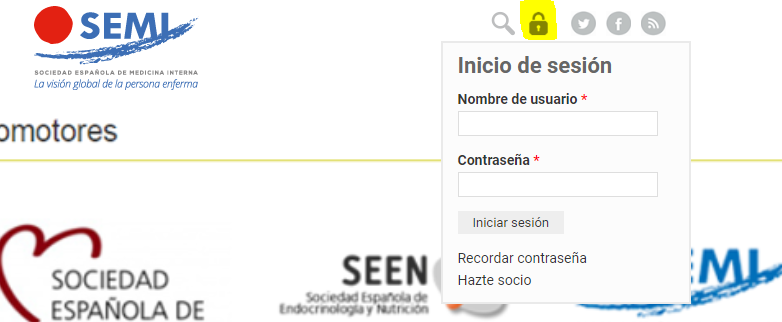
Diríjase desde aquí a la web de la >>>FESEMI<<< e inicie sesión mediante el formulario que se encuentra en la barra superior, pulsando sobre el candado.
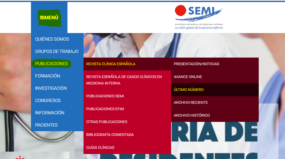
Una vez autentificado, en la misma web de FESEMI, en el menú superior, elija la opción deseada.
>>>FESEMI<<<

