Journal Information
Vol. 214. Issue 9.
Pages 499-504 (December 2014)
Visits
570
Vol. 214. Issue 9.
Pages 499-504 (December 2014)
Original article
Full text access
Clinico-epidemiological characteristics of acute respiratory infections by coronavirus OC43, NL63 and 229E
Características de las infecciones respiratorias agudas causadas por los coronavirus OC43, NL63 y 229E
Visits
570
J. Reina
Corresponding author
jorge.reina@ssib.es

Corresponding author.
, C. López-Causapé, E. Rojo-Molinero, R. Rubio
Unidad de Virología, Servicio de Microbiología, Hospital Universitario Son Espases, Palma de Mallorca, Islas Baleares, Spain
This item has received
Article information
Abstract
Full Text
Bibliography
Download PDF
Statistics
Tables (1)
Table 1. General characteristics of patients with respiratory tract infections in whom coronavirus was detected.
Abstract
Background and objectives

Acute respiratory infection is a very common condition in the general population. The majority of these infections are due to viruses. This study attempted to determine the clinical and epidemiological characteristics of adult patients with respiratory infection by the coronavirus OC43, NL63 and 229E.

Patients and methods

Between January 2013 and February 2014, we prospectively studied all patients with suspected clinical respiratory infection by taking throat swabs and performing a reverse transcription polymerase chain reaction in search of coronavirus.
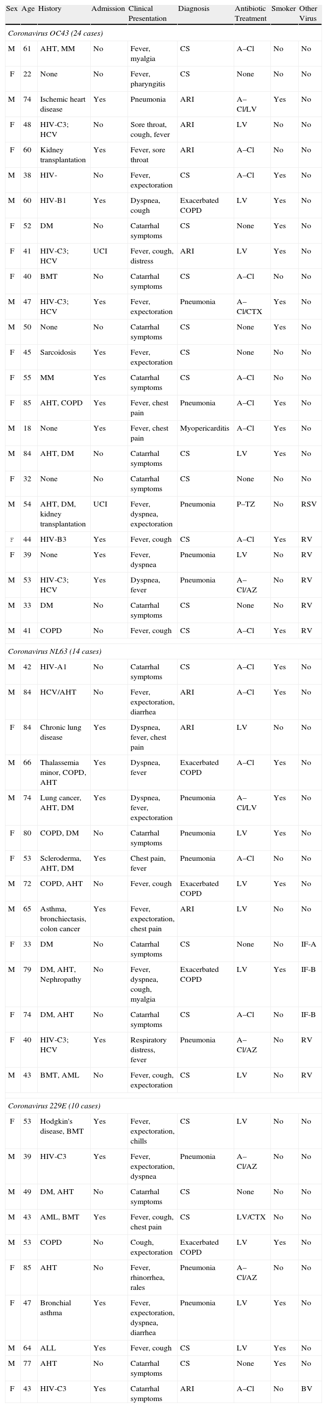
Results

In 48 cases (7.0% of the 686 enrolled patients; 12.6% of the 381 in whom a virus was detected) the presence of a coronavirus demonstrated. In 24 cases, the virus was OC43 (50%); in 14 cases, the virus was NL63 (29%); and in 10 cases, the virus was 229E (21%). The mean age was 54.5 years, with a slight predominance of men. The most common clinical presentations were nonspecific influenza symptoms (43.7%), pneumonia (29.2%) and chronic obstructive pulmonary disease exacerbation (8.3%). Fifty-two percent of the patients required hospitalization, and 2 patients required intensive care. There were no deaths.

Conclusion

Acute respiratory infections caused by coronavirus mainly affect middle-aged male smokers, who are often affected by previous diseases. The most common clinical picture has been nonspecific influenza symptoms.

Keywords:
Coronavirus OC43, NL63, 229E
Acute respiratory infection
Epidemiology
Resumen
Antecedentes y objetivos

Las infecciones respiratorias agudas son una entidad muy frecuente en la población general. La mayoría de ellas son debidas a infecciones víricas. Este estudio pretende precisar las características clínicas y epidemiológicas de los pacientes adultos con infección respiratoria por los coronavirus OC43, NL63 y 229E.

Pacientes y métodos

Entre enero del 2013 y febrero del 2014 se estudió prospectivamente a todos los pacientes con sospecha clínica de infección respiratoria mediante la toma de un frotis faríngeo y la realización de una reacción en cadena de la polimerasa en transcripción reversa en tiempo real en búsqueda de coronavirus.

Resultados

En 48 casos (7,0% de los 686 pacientes incluidos; 12,6% de los 381 en los que se detectó algún virus) se pudo demostrar la presencia de algún coronavirus. En 24 casos se trataba del OC43 (50%), en 14 del NL63 (29%) y en 10 del 229E (21%). La edad media fue de 54,5 años, con un ligero predominio de varones. Las presentaciones clínicas más frecuentes fueron el cuadro gripal inespecífico (43,7%), la neumonía (29,2%) y la agudización de enfermedad pulmonar obstructiva crónica (8,3%). El 52% de los pacientes precisaron ingreso hospitalario, en 2 ocasiones en cuidados intensivos. No se produjo ningún fallecimiento.

Conclusión

Las infecciones respiratorias agudas causadas por coronavirus inciden preferentemente en varones fumadores en la edad media de la vida, frecuentemente afectados de enfermedades previas. La sintomatología clínica mas frecuente ha sido el cuadro gripal inespecífico.

Palabras clave:
Coronavirus OC43, NL63, 229E
Infección respiratoria aguda
Epidemiología
Full Text

What we know?

Respiratory infections are frequently caused by viruses. The use of molecular techniques helps us better understand the etiological spectrum of the viruses involved. This study attempts to determine the involvement of the human coronavirus OC43, NL63 and 2229E in our community.

What this article provides?

The presence of a virus was demonstrated in 55.5% of the included patients, 12.6% of which involved the coronavirus, affecting mostly middle-aged smokers with underlying diseases. The most common clinical presentation was catarrhal syndrome and pneumonia. Fifty-two percent of the patients were hospitalized and had good outcomes, with no case ending in death.

The Editors

Background

Acute respiratory tract infections affect the general population. However, these infections have a higher incidence in the early and late ages of life and in immunosuppressed patients, in whom the infections can be more severe.1 These respiratory infections are frequently caused by viruses, especially the respiratory syncytial virus (RSV) in children and the adenovirus and influenza viruses in adults.2 However, the growing use of molecular diagnostic techniques has broadened the spectrum of viruses involved.2,3

Human coronaviruses are RNA viruses that taxonomically belong to the Coronavirinae subfamily and are classified into 3 distinct genera. The genus Alphacoronavirus includes the coronaviruses 229E and NL63, while the genus Betacoronavirus contains the coronaviruses OC43 and HKU1.1 The latter genus also includes the coronavirus that causes severe acute respiratory syndrome4 and the recently described MERS-CoV, which causes Middle East respiratory syndrome, although its definitive taxonomic position has not yet been established.5

There are few studies that have focused on defining the virus’ role in acute respiratory infections in adults, and those that have analyzed the role of the coronavirus are even scarcer.2,6 We therefore believe that our experience with this type of viral respiratory infection is of interest.

Material and methods

This was a series of prospective cases between January 2013 and February 2014, which included all patients with acute respiratory infections caused by coronavirus in a population over 18 years of age. Throat swabs were taken and assessed for the presence of the coronaviruses OC43, NL63 and 229E. These swabs were used as the only respiratory sample to avoid potential difficulties in the interpretation of the results based on the sample type. The throat swabs were sent to the laboratory in a viral transport medium (MTV, Vircell, Granada, Spain). Only one sample per patient was considered. The samples were processed to detect the various respiratory viruses using a real-time genomic amplification technique (commercial real-time reverse transcription polymerase chain reaction kit), which provides simultaneous and differential detection of 16 different respiratory viruses (Anyplex RV16, Seegene, South Korea). Blood cultures were performed for patients with suspected pneumonia, and serological determinations for Mycoplasma pneumoniae and Chlamydia pneumoniae were conducted. The medical records and epidemiological data of the patients with coronavirus were reviewed. The statistical analysis was performed using the chi-squared test and Student's t-test for paired data. Values of p<.05 were considered significant.

Results

The study took place on the island of Mallorca, with patients with no history of trips abroad in the last 15 days and no known contact with farm or wild animals. A total of 686 patients older than 14 years with suspected viral respiratory infection were included. The presence of a virus was detected in 381 (55.5%) of these patients. In 48 cases (7.0% of the 686 enrolled patients; 12.6% of the 381 in whom a virus was detected), the presence of a coronavirus was demonstrated. In 24 cases, the virus corresponded to OC43 (50%); in 14 cases, the virus was NL63 (29%); and in 10 cases, the virus was 229E (21%). In these 48 patients, the blood cultures were negative, and the serologies did not indicate any other diagnosis.

The main characteristics of each patient with acute human coronavirus infection are listed in Table 1. The mean patient age was 54.5 years (range, 18–85 years); the mean ages of the patients infected by coronavirus OC43, NL63 and 229E were 49 years (range, 18–84 years), 63.5 years (range 33–84 years) and 55.3 years (range 39–85 years), respectively (p>.05). Twenty-six patients (54.1%) were men and 22 (45.9%) were women (p>.05). A predominance of men (60%) was observed in the coronavirus 229E infections. Although cases of coronavirus infection were detected over the entire course of the year, 58.3% of the cases occurred between January and March. In 36 cases (75%), the coronavirus was detected as the only virus, and in 12 cases (25%) it was detected as a coinfection with other viruses (7 cases of rhinovirus, 3 cases of influenza virus, 1 case of RSV and 1 case of bocavirus). The coronavirus 229E had the lowest frequency of mixed infection (10%).

Table 1.

General characteristics of patients with respiratory tract infections in whom coronavirus was detected.

Sex  Age  History  Admission  Clinical Presentation  Diagnosis  Antibiotic Treatment  Smoker  Other Virus 
Coronavirus OC43 (24 cases)
61  AHT, MM  No  Fever, myalgia  CS  A–Cl  No  No 
22  None  No  Fever, pharyngitis  CS  None  No  No 
74  Ischemic heart disease  Yes  Pneumonia  ARI  A–Cl/LV  Yes  No 
48  HIV-C3; HCV  No  Sore throat, cough, fever  ARI  LV  No  No 
60  Kidney transplantation  Yes  Fever, sore throat  ARI  A–Cl  No  No 
38  HIV-  No  Fever, expectoration  CS  A–Cl  Yes  No 
60  HIV-B1  Yes  Dyspnea, cough  Exacerbated COPD  LV  Yes  No 
52  DM  No  Catarrhal symptoms  CS  None  Yes  No 
41  HIV-C3; HCV  UCI  Fever, cough, distress  ARI  LV  Yes  No 
40  BMT  No  Catarrhal symptoms  CS  A–Cl  No  No 
47  HIV-C3; HCV  Yes  Fever, expectoration  Pneumonia  A–Cl/CTX  Yes  No 
50  None  No  Catarrhal symptoms  CS  None  Yes  No 
45  Sarcoidosis  Yes  Fever, expectoration  CS  None  No  No 
55  MM  Yes  Catarrhal symptoms  CS  A–Cl  No  No 
85  AHT, COPD  Yes  Fever, chest pain  Pneumonia  A–Cl  Yes  No 
18  None  Yes  Fever, chest pain  Myopericarditis  A–Cl  Yes  No 
84  AHT, DM  No  Catarrhal symptoms  CS  LV  Yes  No 
32  None  No  Catarrhal symptoms  CS  None  No  No 
54  AHT, DM, kidney transplantation  UCI  Fever, dyspnea, expectoration  Pneumonia  P–TZ  No  RSV 
F  44  HIV-B3  Yes  Fever, cough  CS  A–Cl  Yes  RV 
39  None  Yes  Fever, dyspnea  Pneumonia  LV  No  RV 
53  HIV-C3; HCV  Yes  Dyspnea, fever  Pneumonia  A–Cl/AZ  No  RV 
33  DM  No  Catarrhal symptoms  CS  None  No  RV 
41  COPD  No  Fever, cough  CS  A–Cl  Yes  RV 
Coronavirus NL63 (14 cases)
42  HIV-A1  No  Catarrhal symptoms  CS  A–Cl  Yes  No 
84  HCV/AHT  No  Fever, expectoration, diarrhea  ARI  A–Cl  Yes  No 
84  Chronic lung disease  Yes  Dyspnea, fever, chest pain  ARI  LV  No  No 
66  Thalassemia minor, COPD, AHT  Yes  Dyspnea, fever  Exacerbated COPD  A–Cl  Yes  No 
74  Lung cancer, AHT, DM  Yes  Dyspnea, fever, expectoration  Pneumonia  A–Cl/LV  Yes  No 
80  COPD, DM  No  Catarrhal symptoms  Pneumonia  LV  Yes  No 
53  Scleroderma, AHT, DM  Yes  Chest pain, fever  Pneumonia  A–Cl  No  No 
72  COPD, AHT  No  Fever, cough  Exacerbated COPD  LV  Yes  No 
65  Asthma, bronchiectasis, colon cancer  Yes  Fever, expectoration, chest pain  ARI  LV  No  No 
33  DM  No  Catarrhal symptoms  CS  None  No  IF-A 
79  DM, AHT, Nephropathy  No  Fever, dyspnea, cough, myalgia  Exacerbated COPD  LV  Yes  IF-B 
74  DM, AHT  No  Catarrhal symptoms  CS  A–Cl  No  IF-B 
40  HIV-C3; HCV  Yes  Respiratory distress, fever  Pneumonia  A–Cl/AZ  No  RV 
43  BMT, AML  No  Fever, cough, expectoration  CS  LV  No  RV 
Coronavirus 229E (10 cases)
53  Hodgkin's disease, BMT  Yes  Fever, expectoration, chills  CS  LV  No  No 
39  HIV-C3  Yes  Fever, expectoration, dyspnea  Pneumonia  A–Cl/AZ  No  No 
49  DM, AHT  No  Catarrhal symptoms  CS  None  No  No 
43  AML, BMT  Yes  Fever, cough, chest pain  CS  LV/CTX  No  No 
53  COPD  No  Cough, expectoration  Exacerbated COPD  LV  Yes  No 
85  AHT  No  Fever, rhinorrhea, rales  Pneumonia  A–Cl/AZ  No  No 
47  Bronchial asthma  Yes  Fever, expectoration, dyspnea, diarrhea  Pneumonia  LV  Yes  No 
64  ALL  Yes  Fever, cough  CS  LV  Yes  No 
77  AHT  No  Catarrhal symptoms  CS  None  Yes  No 
43  HIV-C3  Yes  Catarrhal symptoms  ARI  A–Cl  No  BV 

General characteristics of the patients enrolled in this study with respiratory tract infections in whom the presence of coronavirus OC43, NL63 or 229E was detected.

A–Cl: amoxicillin–clavulanate; AHT: arterial hypertension; ALL: acute lymphoblastic leukemia; AML: acute myeloid leukemia; ARI: acute lower respiratory tract infection; AZ: azithromycin; BMT: bone marrow transplantation; BV: bocavirus; CA: cancer; COPD: chronic obstructive pulmonary disease; CS: catarrhal syndrome; CTX: cotrimoxazole; DM: diabetes mellitus; F: female; HCV: liver disease due to hepatitis C virus infection; HIV: human immunodeficiency virus; IF: influenza; LV: levofloxacin; M: male; MM: multiple myeloma; P–TZ: piperacillin–tazobactam; RSV: respiratory syncytial virus; RV: rhinovirus.

Twenty-three patients (47.9%) were smokers. Only 15 (31.2%) had been vaccinated for seasonal influenza. Eleven patients (22.9%) were infected with the human immunodeficiency virus (HIV). The main respiratory syndromes associated with the coronaviruses were catarrhal syndromes (fever, general discomfort, pharyngitis, adynamia) (occurring in 21 patients, 43.7%), pneumonia (occurring in 14 patients, 29.1%) and exacerbation of chronic obstructive pulmonary disease (COPD) (occurring in 4 patients, 8.3%). Twenty-five patients (52%) were hospitalized, 2 of them (8%) in the intensive care unit. Thirty-nine patients (81.2%) underwent antibiotic treatment (21 patients were treated with amoxicillin/amoxicillin, and 18 were treated with levofloxacin), which typically was maintained despite the viral diagnosis. There were no deaths.

Discussion

Acute respiratory infections caused by coronavirus in the general population represent 2–18% of cases, depending on the geographical area, type of population studied and viral detection technique employed.7 In a large 4-year study performed in France, Lepiller et al.8 found that coronaviruses represented 11.7% of the analyzed cases. However, in Brazil, these viruses only represented 1.2% of the cases studied from 2009 to 2010,9 while in China, the rates were 4.4%10 and 11.7%11 for a pediatric population and an adult population, respectively. In a study performed in Canada,12 coronaviruses only represented 2% of the respiratory samples from adults, which is consistent with that observed by Gaunt et al.13 in Great Britain (2.3%). However, all of these estimates generally refer to the rate of positivity for a total number of samples analyzed. Thus for the Gaunt et al. study,13 the rate of etiologic participation of the coronaviruses rose to 8.1% of all the samples positive to any virus. In our study, we detected coronaviruses in 6.9% of the enrolled patients and in 12.6% of those in whom a virus was demonstrated, with readings very close to those measured in neighboring countries, such as France.8

The distribution of the various types of coronavirus varied widely according to the geographical area. Thus, while only NL63 and OC43 were detected in Brazil,9 Lepiller et al.8 observed a predominance in France of the coronavirus 229E (53%) compared to the others (36% for NL63 and 11% for OC43). However, Gaunt et al.13 observed in their series of 221 cases in Edinburgh that the most common coronavirus was OC43 (50.2%), followed by NL63 (33.9%) and 229E (15.8%). In our study, which was shorter and had fewer cases of infections, the coronavirus OC43 (50%) was also predominant, followed by NL63 (29%) and 229E (21%).

One of the common characteristics of most respiratory viruses is that they usually cause mixed infections, in conjunction with other respiratory viruses.14 The rates of coinfection vary from 30 to 70%3,14; patients with mixed infections do not have higher rates of morbidity or mortality.8,13 The detection of probable mixed infections should be assessed with caution because they can reflect respiratory infections prior to the acute process that motivated the study. The simultaneous detection of several viruses through genomic tests also establishes the possibility that some patients are carriers of any of the detected viruses without them having a direct pathogenic role in the current infection.3,14

In our study, the rate of coinfection was not very high (25%). Gaunt et al.13 reported a 35% coinfection rate, with rhinovirus the most frequent companions (65%); in our study, this virus represented 58.3%. Coinfections with the influenza virus have been described mainly in children, representing 5–10% of the cases.10 Twenty-five percent of coinfection cases in our series corresponded to the influenza virus, probably coinciding with the seasonal flu epidemic. The detection of a case of RSV coinfection is noteworthy, given that a previous study on RSV did not detect any cases of this type of coinfection.15

Although they can be detected over the course of the year, the incidence of coronavirus infections adjusts to a seasonal pattern, occurring predominantly in winter months.2,6,13 A number of studies have observed that OC43 presents a pattern with a biannual variation, with a predominance in the first quarter of the year.13,16 We detected the incidence of respiratory infections by coronavirus over the course of the year, although the majority (58.3%) occurred between the months of January and March.

The mean age of our patients was 54.5 years, with a range of 18–85 years and the infections mainly affecting middle-aged individuals. There are few studies that reflect middle age in adult patients. Bastien et al.12 measured a mean age of 72 years for patients infected by NL63. We observed no significant variations in patient age depending on the type of coronavirus, although the mean age of the patients with NL63 was somewhat higher (63.5 years). We also observed no significant differences in terms of the sex of the patients with infections; however, Gaunt et al.13 reported a somewhat higher rate in men infected by OC43 and NL63.

The coronavirus, as with most other respiratory viruses, causes acute infections in both the upper and lower respiratory tract; the clinical spectrum is therefore difficult to differentiate from other viruses.2,8,13,17 In our study, as in previous studies, the most common respiratory disease was nonspecific catarrhal syndrome.3,8,13,16,18

Pneumonia constitutes another of the typical clinical presentations of coronaviruses; thus, 10–18% rates have been reported.19,20 Lepiller et al.8 found that the clinical presentation of coronavirus infection was pneumonia in 14% of their patients, with values of 26% for 229E, 25% for OC43 and 0% for NL63. In our series, 29.1% of the patients were diagnosed with pneumonia, with a greater rate in infections from coronavirus 229E (40%).

Coronavirus 229E has been described as the predominant causal agent in immunosuppressed patients.21 Eleven patients in our study (22.9%) were infected with HIV; coronavirus 229E was involved as the pathogen in only 2 cases (18%), while OC43 was involved in 7 (64%) and NL63 in 2 (18%). We have found no data referring to the potential implications of coronavirus infections in patients with HIV infection.

Respiratory virus infections can also cause COPD exacerbations, although the coronavirus appears to be less involved than other viruses.8,13,16 Nevertheless, our study detected NL63 in 14.2% of the infected patients, and Lepiller et al.8 found them to be involved in 6% of their patients. These viruses should therefore be considered potential etiologic agents in these processes.

Most of the patients enrolled in our series had an underlying disease (43 patients, 89.6%), which might explain the high rate of empiric antibiotic treatment, which was employed for 38 patients (81.2%), mainly amoxicillin/clavulanic acid (21 patients, 53.8%) and levofloxacin (18 patients, 46.1%). Sixty-six percent of the patients underwent antibiotic treatment even after the presence of coronavirus was detected. At present, there is no antiviral drug that has been proven effective against the human coronavirus.7

Smoking is one of the main predisposing factors for numerous respiratory diseases and, among others, viral infections. Twenty-three of our patients (47.9%) were smokers. Only 15 of the patients in our series (31.2%) had been vaccinated against seasonal influenza.

The hospitalization of patients with acute respiratory infection often depends on the symptoms and severity of the respiratory clinical picture or of the patient's underlying disease. Twenty-five of the patients (52%) in our series were hospitalized, but only 2 (4%) were admitted to the ICU, 1 of whom was a patient infected with HIV and in respiratory distress and the other was a patient with a kidney transplant and bilateral pneumonia. Both of the patients had favorable outcomes. Of the 50 cases described by Lepiller et al.,8 4% were admitted to the ICU, consisting of a case of NL63 infection and a case of 229E infection. Our 2 cases were infected by the coronavirus OC43 (8.3% of the total). As with the study by Lepiller et al.,8 none of our patients died during the coronavirus infection episode.

We can conclude that acute respiratory infections by human coronavirus mostly affect middle-aged men who smoke and have underlying diseases. The predominant clinical presentation is a lower respiratory tract infection, with general malaise, which usually has a favorable outcome.

Conflicts of interest

The authors declare that they have no conflicts of interest.

References
[1]
P.S. Masters, S. Perlman.
Coronaviridae.
Fields virology, 6th ed., pp. 825-858
[2]
H.K. Talbot, A.R. Falsey.
The diagnosis of viral respiratory disease in older adults.
Clin Infect Dis, 50 (2010), pp. 747-751
[3]
L.J. Van Elden, A.M. Van Loon, F. Van Alphen, K.A. Hendriksen, A.I. Hoepelman, M.G. Van Kraaij, et al.
Frequent detection of human coronaviruses in clinical specimens from patients with respiratory tract infection by use of a novel real-time reverse-transcriptase polymerase chain reaction.
J Infect Dis, 189 (2004), pp. 652-657
[4]
J.S. Peiris, K.Y. Yuen, A.D.M.E. Osterhaus, K. Stöhr.
The severe acute respiratory syndrome.
N Engl J Med, 349 (2003), pp. 2431-2441
[5]
A.M. Zaki, S. Van Boheemen, T.M. Besterbroer, A.D.M.E. Osterhaus, R.A. Fouchier.
Isolation of a novel coronavirus from a man with pneumonia in Saudi Arabia.
N Engl J Med, 367 (2012), pp. 1814-1820
[6]
A.R. Falsey, E.E. Walsh, F.G. Hayden.
Rhinovirus and coronavirus infection-associated hospitalization among older adults.
J Infect Dis, 185 (2002), pp. 1338-1341
[7]
S.B. Greenberg.
Update on rhinovirus and coronavirus infections.
Sem Res Crit Care Med, 32 (2011), pp. 433-446
[8]
Q. Lepiller, H. Barth, F. Lefebvre, R. Herbrecht, P. Lutz, R. Kessler, et al.
High incidence but low burden of coronaviruses and preferential associations between respiratory viruses.
J Clin Microbiol, 51 (2013), pp. 3039-3046
[9]
T.K. Cabeça, E. Carraro, A. Watanabe, C. Granato, N. Bellei.
Infections with human coronaviruses NL63 and OC43 among hospitalised and outpatient individuals in Sao Paulo, Brazil.
Mem Inst Oswaldo Cruz, 107 (2012), pp. 693-694
[10]
S.S. Chiu, K.H. Chan, K.W. Chu, S.W. Kwan, Y. Guan, L.L. Poon, et al.
Human coronavirus NL63 infection and other coronavirus infections in children hospitalized with acute respiratory disease in Hong Kong, China.
Clin Infect Dis, (2005), pp. 1721-1729
[11]
X. Yu, R. Lu, Z. Wang, N. Zhu, W. Wang, D. Julian, et al.
Etiology and clinical characterization of respiratory virus infections in adult patients attending an emergency department in Beijing.
[12]
N. Bastien, K. Anderson, L. Hart, P. Van Caeseele, K. Brandt, D. Milley, et al.
Human coronavirus NL63 infection in Canada.
J Infect Dis, 191 (2005), pp. 503-506
[13]
E.R. Gaunt, A. Hardie, E.C.J. Claas, P. Simmonds, K.E. Templeton.
Epidemiology and clinical presentations of the four human coronaviruses 229E, HKU1, NL63 and OC43 detected over 3 years using a novel multiplex real-time PCR method.
J Clin Microbiol, 48 (2010), pp. 2940-2947
[14]
L. Ren, R. Gonzalez, Z. Wang, Z. Xiang, Y. Wang, H. Zhou, et al.
Prevalence of human respiratory viruses in adults with acute respiratory tract infections in Beijing, 2005–2007.
Clin Microbiol Infect, 15 (2009), pp. 1146-1153
[15]
J. Reina, C. López.
Infecciones respiratorias causadas por el virus respiratorio sincitial en la población adulta: descripción de una serie de 16 casos.
Med Clin Barc, 141 (2013), pp. 163-166
[16]
G. Gerna, E. Percivalle, A. Sarasini, G. Campanini, A. Piralta, F. Rovida, et al.
Human respiratory coronavirus HKU1 versus other coronavirus infections in Italian hospitalised patients.
J Clin Virol, 38 (2007), pp. 244-250
[17]
S. Lau, Y. Woo, Y. Yip, H. Tse, W. Tsoi, C. Cheng, et al.
Coronavirus HKU1 and other coronavirus infections in Hong Kong.
J Clin Microbiol, 44 (2006), pp. 2063-2071
[18]
R. Njouom, E.L. Yekwa, P. Cappy, A. Vabret, P. Boisier, D. Rosset.
Viral etiology of influenza-like illness in Cameroon, January–December 2009.
J Infect Dis, 206 (2012), pp. S29-S35
[19]
L.C. Jennings, T.P. Anderson, K.A. Beynon, A. Chua, R.T. Laing, A.M. Werno, et al.
Incidence and characteristics of viral community-acquired pneumonia in adults.
Thorax, 63 (2008), pp. 42-48
[20]
D. Lieberman, A. Shimoni, Y. Shemer-Avni, A. Keren-Naos, R. Shtainberg.
Respiratory viruses in adults with community-acquired pneumonia.
Chest, 138 (2010), pp. 811-816
[21]
F. Pene, A. Merlat, A. Vabret, F. Rozenberg, A. Buzyn, F. Dreyfus, et al.
Coronavirus 229E related pneumonia in immunocompromised patients.
Clin Infect Dis, 37 (2003), pp. 929-932

Please cite this article as: Reina J, López-Causapé C, Rojo-Molinero E, Rubio R. Características de las infecciones respiratorias agudas causadas por los coronavirus OC43, NL63 y 229E. Rev Clin Esp. 2014;214:499–504.

Copyright © 2014. Elsevier España, S.L.U.. All rights reserved
Idiomas
Revista Clínica Española (English Edition)
Article options
Tools
es en

¿Es usted profesional sanitario apto para prescribir o dispensar medicamentos?

Are you a health professional able to prescribe or dispense drugs?