Journal Information
Share
Share
Download PDF
More article options
Visits
147
Review
Full text access
Uncorrected Proof. Available online 4 December 2024
Potential role of lung ultrasonography in outpatient follow-up of patients with COVID-19. A systematic review
Papel de la ecografía pulmonar en el seguimiento ambulatorio de pacientes tras COVID-19. Revisión sistemática de la literatura
Visits
147
F.N. Romeroa,b,
Corresponding author
f.navarroromero@gmail.com

Corresponding author.
, J.O. Sierraa, M.D. Martín Escalantea
a Servicio de Medicina Interna, Hospital Costa del Sol, 29603 Málaga, Spain
b Facultad de Medicina, Universidad de Málaga, 29010 Málaga, Spain
This item has received
Article information
Statistics
Figures (1)
Tables (2)
Table 1. Thoracic ultrasound characteristics and results in published studies.
Table 2. Quality of the ultrasound technique and operator observation.
Show moreShow less
Abstract
Introduction and aim

Currently, the usefulness of lung ultrasound in the follow-up of patients after hospital discharge for SARS-CoV-2 pneumonia is not well known. The main objective of this systematic review is to investigate the persistence of alterations in lung ultrasound of patients who have had COVID-19 pneumonia.

Methods

A systematic review has been carried out following the PRISMA regulations in the PubMed, EMBASE, Web of Science and Google Scholar database from January 2020 to May 2023 using the combination of MeSH terms: “lung ultrasound”, “ultrasonography”, “lung alterations”, “persistence”, “follow-up”, “consequences”, “hospital discharge”, “COVID”, “COVID-19”, “SARS-CoV-2”. Studies were selected that described alterations in the lung ultrasound of patients after having suffered from COVID-19 pneumonia. The JBI Critical Appraisal Tools were used to assess the risk of bias of the studies. No meta-analysis techniques were performed, the results being compared narratively.

Results

From two to six months after COVID-19 pneumonia, pulmonary ultrasound abnormalities appear frequently and are proportional to the intensity of the initial episode. The most frequent anomalies are irregularities in the pleural line, the presence of B lines and/or subpleural consolidations, predominantly in the basal regions of the thorax. These findings seem to correlate with those of the chest CT.

Conclusions

Lung ultrasound offers technical and economic advantages that should be considered for the study of patients after hospital discharge for COVID-19.

Keywords:
Lung ultrasound
SARS-CoV-2
COVID-19
Consequences
Abbreviations:
WHO
ARDS
CT
ICU
ILD
IMV
NIMV
Resumen
Introducción y objetivo

Actualmente no se conoce bien la utilidad de la ecografía pulmonar en el seguimiento de los pacientes tras haber tenido una hospitalización por neumonía por SARS-CoV-2. El objetivo principal de esta revisión sistemática es investigar la persistencia de las alteraciones en la ecografía pulmonar de los pacientes que han tenido una neumonía COVID-19.

Métodos

Se ha realizado una revisión sistemática siguiendo la normativa PRISMA en la base de datos PubMed, EMBASE, Web os Science y Google Scholar desde enero del 2020 hasta mayo del 2023 utilizando la combinación de términos MeSH: lung ultrasound, ultrasonography, lung alterations, persistence, follow-up, consequences, hospital discharge, COVID, COVID-19, SARS-CoV-2. Se seleccionaron estudios que describieran alteraciones en la ecografía pulmonar de pacientes tras haber padecido una neumonía COVID-19. Se empleó la JBI Critical Appraisal Tools para evaluar el riesgo de sesgos de los estudios. No se realizaron técnicas de metaanálisis, siendo los resultados comparados narrativamente.

Resultados

De dos a seis meses después de la neumonía COVID-19, las alteraciones ecográficas pulmonares aparecen con frecuencia y son proporcionales a la intensidad del episodio inicial. Las anomalías más frecuentes son las irregularidades de la línea pleural, la presencia de líneas B y/o consolidaciones subpleurales, con predominio por las regiones basales del tórax. Estos hallazgos parecen correlacionarse con los de TC de tórax.

Conclusiones

La ecografía pulmonar ofrece unas ventajas técnicas y económicas que deben considerarse para el estudio de los pacientes tras el alta hospitalaria por COVID-19.

Palabras clave:
Ecografía pulmonar
SARS-CoV-2
COVID-19
Consecuencias
Full Text
Introduction

SARS-CoV-2 disease (COVID-19) has posed a threat to global health since its emergence. The first case of the disease was declared in the city of Wuhan in late December 2019, with rapid transmission of the virus to the rest of the world.1 The World Health Organization (WHO) declared a pandemic on March 11, 2020.2 As of May 2023, more than 766 million cases had been reported with more than 6.9 million deaths had resulted from the disease.3

The main organ affected by COVID-19 is the lung. The clinical spectrum ranges from an asymptomatic carrier to atypical pneumonia and the possibility of developing ARDS.1,4–6

Various studies have shown persistent symptoms after acute infection. The most prevalent respiratory symptoms are dyspnea and cough. A systematic literature review found that six months after the acute episode, dyspnea was present in 29.7% (CI 14.9%–44.0%) and cough in 13.1% (CI 5.3%–22.6%).7

Guidelines and study protocols on patient follow-up have been developed by different scientific societies. Follow-up with a chest x-ray and/or chest CT scan is suggested, but to date, the role of thoracic ultrasound in this scenario has not been fully evaluated.8,9

Chest x-ray is not sensitive enough to detect pulmonary changes in acute infection. Its most frequent findings are interstitial infiltrates and consolidations.10,11 A chest CT scan is considered the gold standard test. It has a higher sensitivity for detecting pneumonia than a chest x-ray.12,13 The most frequent and earliest pattern on a CT scan is ground glass, which is usually multifocal, bilateral, and peripheral.14 As the pneumonia progresses, the affected areas may grow and develop a cobblestone pattern.15 Some studies performed after hospital discharge highlight the persistence of pulmonary abnormalities. At four weeks after discharge, the most frequent findings in 51 patients with COVID-19 pneumonia were thickened interlobular septa and ground glass patterns (54%).16 In a six-month study of 114 patients with severe pneumonia, ground glass patterns were observed in 21% and fibrotic-like changes (parenchymal bands, traction bronchiectasis, pannalization) in 35%.17

Lung ultrasound proved useful during the COVID-19 pandemic. A technique known in the clinical setting since the early 1990s, its use in COVID-19 infection was notable since the beginning of the pandemic and grew exponentially.18 In COVID-19, it was used for screening and follow-up on patients during the acute phase of the disease in emergency departments, hospital wards, and ICUs.19–21 However, little is known about its usefulness in the follow-up of patients after hospital discharge.

As a summary of the main findings on lung ultrasound during acute SARS-CoV-2 infection, pleural line irregularities and B-lines with a heterogeneous appearance appear initially. As disease severity increases, B-lines increase and subpleural lung consolidations appear. With progression to ARDS, so-called "white lung" is caused by pulmonary edema.21,22

Thoracic ultrasound has the advantage of being a quick test that is widely available, is able to be performed at the patient’s bedside, is economical, does not use ionizing radiation, and has a fast learning curve.12,23 It is appropriate to review its involvement after acute COVID-19 infection.

Methods

A systematic review was performed based on the criteria of the Preferred Reporting Items for Systematic Reviews and Meta-Analyses (PRISMA) statement list.24

The research question was: In patients who have been hospitalized for COVID-19 pneumonia, does performing a lung ultrasound at least 60 days after hospital discharge detect abnormalities?

A literature search was performed in the PubMed, EMBASE, Web of Science, and Google Scholar databases, collecting studies published from January 2020 to May 2023 with the MeSH terms: [(lung ultrasound OR ultrasonography OR lung alterations) AND (persistence OR follow-up OR consequences OR hospital discharge) AND (COVID OR COVID-19 OR SARS-CoV-2)].

The inclusion criteria were:

  • -

    Studies of patients ≥ 18 years with follow-up after hospital discharge for SARS-CoV-2 pneumonia.

  • -

    In the study, a thoracic ultrasound must have been performed ≥60 days after hospital discharge.

  • -

    The study must have recorded the ultrasound signs in a systematic manner and by chest segments.

The exclusion criteria were:

  • -

    Age < 18 years.

  • -

    Studies that do not include a pulmonary ultrasound in the follow-up.

Article selection was determined using the consecutive phases of the PRISMA method. The main variables extracted were the thoracic ultrasound modality and the ultrasound signs described (pleural line irregularities, B-lines, subpleural consolidations). Secondary variables were age, sex, country, study period, sample size, comorbidity (hypertension, dyslipidemia, obesity), and need for ventilatory support. The most relevant data were collected and compared in a table (as synthesis techniques were not used in the study).

In the eligibility phase, three reviewers (FNR, JOS, MDME) read the full text of the articles, evaluated them independently, and subsequently extracted the data. The data extracted from the studies were verified by each reviewer. It was unanimously decided to include the data in a table comparing the results for a qualitative analysis. The quality of the method used and the risk of bias in the articles was evaluated with the JBI Critical Appraisal Tools25 for observational studies and were assessed individually by the three reviewers. The study was considered adequate if the frequency of ultrasound signs was collected in a systematized way. In studies that did not describe the frequency of ultrasound signs, the appendices were consulted and if these data were not available, they were not included in the analysis. Meta-analysis techniques were not performed; the results were compared in a narrative. The study protocol can be consulted in the Open Science Framework at https://doi.org/10.17605/OSF.IO/8QRM5.

Results

The flow chart of the literature search and final selection of articles is shown in Fig. 1.

Figure 1.

Flow chart of the study selection process (PRISMA).24

(0.3MB).
Findings of the studies included

Giovannetti et al.26 was the first study to evaluate the role of lung ultrasound in the long-term follow-up of patients. It also compared lung ultrasound findings with a chest CT scan performed 90 days after hospital discharge. Three months after discharge, subjects had a 12-region lung ultrasound and a thoracic CT scan. Thirty-eight patients were included. The mean age was 60 years and 89.5% had comorbidities (hypertension 55.3%, dyslipidemia 21.1%, obesity 18.4%). A total of 68.4% required CPAP, 21.1% high-flow nasal cannula therapy, and 10.5% BiPAP. None were admitted to the ICU.

For each of the thoracic regions, a score was given according to the LUS score27: 0 points: normal pattern (A-lines or <3 B-lines); 1 point: moderate pleural line loss or ≥3 B-lines; 2 points: severe pleural line loss or coalescing B-lines; 3 points: complete pleural line loss or white lung and/or lung consolidations. According to the score, lung ultrasound involvement was classified as mild (LUS score between 1 and 7 points), moderate (between 8 and 18 points), or severe (between 19 and 36 points).

At 90 days, 63.2% of subjects had B-lines suggestive of residual interstitial lung disease. The involvement was mild in 42.1%, moderate in 21.1%, and severe in no subjects.

This study was the first to correlate lung ultrasound findings with a chest CT scan during follow-up after discharge for COVID-19 pneumonia. Findings suggestive of an interstitial pattern on the lung ultrasound were confirmed on a chest CT scan. A concordance analysis of both tests showed a ROC curve of 0.94 with good discriminatory ability of the lung ultrasound and a Cohen’s kappa coefficient of 0.74, with a good concordance rate between both tests.

Alharthy et al.28 analyzed lung ultrasound findings at the time of hospital admission to the ICU and followed up on pulmonary ultrasound findings at two and four months after hospital discharge. A total of 171 patients were included. The mean age was 47 years and 78.9% were men. A total of 69.6% required IMV and the remaining 30.4% used high-flow nasal cannula therapy, CPAP, or a Venturi mask.

They performed a lung ultrasound of the 12 thoracic regions. On the analysis at ICU admission, there was a high prevalence of pulmonary ultrasound abnormalities that were significantly greater in non-survivors compared to survivors.

In the analysis of the lung ultrasound at two and four months after hospital discharge, ultrasound abnormalities continued to be found, with the presence of B-lines, pleural line irregularities, and pulmonary consolidations. At two months, there was a predominance of B-line involvement in the thoracic bases (40.2% in the posterior inferior region and 43.3% with bilateral involvement), irregularities of the pleural line in more than six areas in 52.5%, and presence of pulmonary condensations in 44.9% in the posterior inferior region (31.5% bilaterally). At four months, lung ultrasound abnormalities continued to persist, but at a lower frequency than at two months. At four months, 22.8% had B-lines in the posterior inferior region and 26.8% in the anterior inferior region, 22.5% had pleural line irregularities in more than six areas, and 32.3% had pulmonary consolidations in the posterior inferior region (18.9% bilaterally).

Hernández-Píriz et al.29 evaluated patients with a 13-region lung ultrasound during hospitalization, at one month, and at three months after discharge. Ninety-six patients were included. The mean age was 55 years and 55.2% were men. A total of 56.2% were obese, 30.2% hypertensive, and 10.4% diabetic. A total of 77% had ARDS and 6.2% required IMV.

At one month after discharge, 82.5% of the cases had ultrasound abnormalities in the posterior inferior region, 74.2% in the anterior region, and 80.2% had bilateral pulmonary involvement. At three months after discharge, 68.7% of patients had improved lung ultrasound signs. The posterior inferior region continued to be the most affected region (30.9%), followed by the anterior region (26.8%). Bilateral pulmonary involvement persisted in 31.2%. For the subgroup of patients with moderate and severe ARDS, the persistence of lung ultrasound lesions was higher and 32.4% and 61.5% had bilateral involvement, respectively, at three months.

Fortini et al.30 evaluated patients three to six months after hospital discharge. The median time to lung ultrasound after discharge was 123 days. Fifty-nine patients were included. The mean age was 68.2 years and 52.5% were men. The most prevalent comorbidity was hypertension (47.5%) followed by cardiovascular disease (30.1%) and obesity (16.9%). No patient had IMV or ICU admission. Twenty-seven percent required some type of non-invasive ventilation (NIV).

A thoracic ultrasound of the 12 lung regions was performed. According to the ultrasound findings, involvement was classified as mild (1–12), moderate (13–24), or severe (25–36). Thoracic ultrasound showed abnormalities in 15 of the 53 patients (28.3%), with findings of B-lines. The involvement was scored as mild in all cases.

Lopes et al.31 conducted a study of patients in post-COVID-19 follow-up with a lung ultrasound evaluation at two and five months after presenting with pneumonia. Fifty-nine patients who had required hospitalization were included. The mean age was 59 years and 54% were women. A total of 32.2% had hypertension and 20.3% were diabetic. A total of 11.9% were admitted to the ICU.

A 12-region thoracic ultrasound was performed. At two months, 78% of subjects had lung ultrasound abnormalities; at five months, 61% had abnormalities. At two months, the most frequent ultrasound abnormality was the presence of >2 B-lines (69.5%), followed by coalescent B-lines (22%), and subpleural consolidations (15.3%). At five months, 57.8% of the subjects continued to have B-lines, 20.3% coalescent B-lines, and 10.2% subpleural consolidations.

Ferioli et al.32 conducted a study of patients after hospitalization, with lung ultrasound performed two months after discharge. A total of 100 patients were included. The mean age was 60 years, 55% were male, and 50% had some cardiovascular comorbidity (hypertension, diabetes, heart disease). A total of 34% required some type of ventilatory support (10 patients IMV and 24 NIV, CPAP, or high-flow nasal cannula therapy); the greater prevalence of cardiovascular comorbidities in this group was significant (74%). Of the 100 study participants, a 12-region chest ultrasound was performed two months after hospital discharge in 53 patients.

The ultrasound abnormality most frequently found was pleural line irregularity, present in 94% of patients, together with associated B-lines.

The study defined interstitial lung disease according to ultrasound as the presence of ultrasound findings in two or more thoracic regions with a LUS score ≥2.33 It was found in 59% of patients.

Gurbani et al.34 conducted a study of patients four months after hospital discharge. Seventy-seven patients were included. The mean age was 57 years; 48% were women; and comorbidities included hypertension (48%), dyslipidemia (30%), diabetes (17%), and obesity (14%). Sixty-four percent required some oxygen therapy support during hospitalization and 14% were admitted to the ICU for IMV.

A 12-region lung ultrasound was performed and 76.5% had B-lines.

Russo et al.35 conducted a study six months after hospital discharge. Seventy-four patients were included. The mean age was 65 years; 73% were men; and comorbidities included hypertension (43%), diabetes (11.8%), and coronary artery disease (6.7%). Fifty percent had severe disease, with NIV in 35 cases (47.3%) and ICU admission in two cases (2.7%).

Of the study subjects, 69.4% had abnormalities on the lung ultrasound, with irregular pleural line in 52.8% and bilateral involvement in 41.9%. The inferior thoracic regions were the most affected (31.8%). Small subpleural condensations (≤1 cm) were observed in 31 patients (43.1%) and all larger condensations had disappeared.

Clofent et al.36 performed a study in a period between two and five months after hospital discharge with a chest CT scan and 12-region lung ultrasound with no more than 15 days between the two tests.

A total of 352 patients were included. The mean age was 56 years; 57.7% were men; and comorbidities included hypertension (35.8%), dyslipidemia (22.4%), diabetes (15.6%), and obesity (33.8%). Eighty-one patients (23%) had critical pneumonia requiring IMV, 51 patients (14.5%) had severe pneumonia with NIV or high-flow nasal cannula therapy, 109 patients (31%) had moderate pneumonia with low-flow nasal cannula therapy, and 111 patients (31.5%) had mild pneumonia without the need for oxygen therapy.

The Warrick scale,37 with a score from 0 to 30, was applied to the chest CT results. A cutoff point ≥7 had previously been validated as a predictor of interstitial lung disease. In this study, patients with a Warrick scale score ≥7 were classified as having relevant interstitial lung sequelae (RILS). For lung ultrasound results, a scale (LUS) was applied according to the findings, adding one point for each thoracic region in which pathological B-lines appear for a total score that ranged from 0 to 12. The scores obtained in both scales—Warrick and LUS—were correlated.

In 257 patients (73%), the presence of B-lines was observed on lung ultrasound, more frequently in the posterior and inferior regions. B-lines were significantly higher in the RILS group compared to the non-RILS group (98.7% vs. 53%, P < .001). The LUS score was significantly higher in the RILS group compared to the non-RILS group (5.0 vs. 1.0, P < .001). The ROC curve showed that a B-line score ≥3 was the best cutoff point for discriminating patients with RILS (sensitivity 94.2%, specificity 81.8%, negative predictive value 94.7%).

Pleural line irregularities were observed in 190 patients (53.9%) and were significantly more common in the RILS group compared to the non-RILS group (77.3% vs. 35.9%, P < .001). Subpleural consolidations were observed in 13 patients (3.7%).

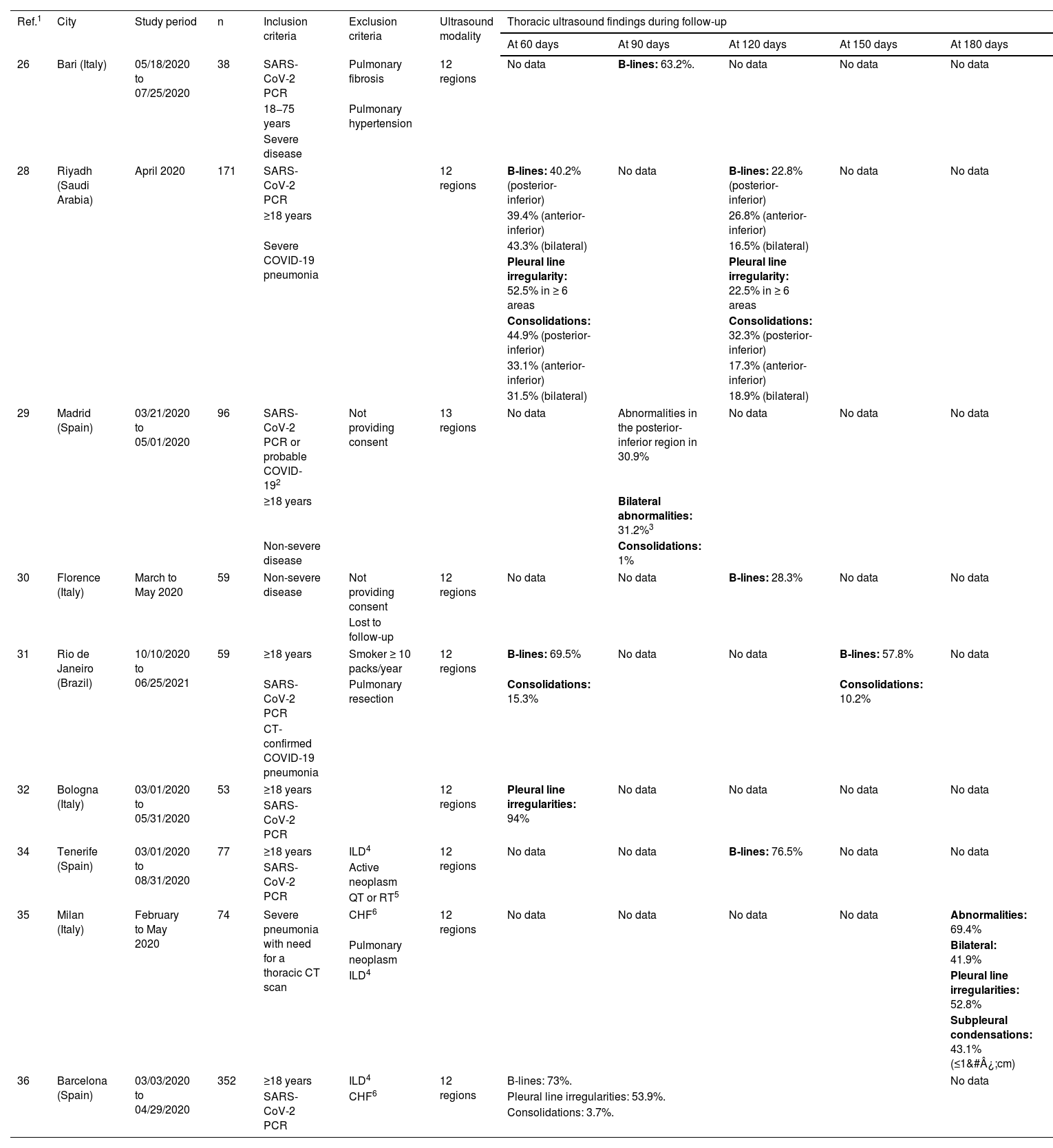
Table 1 describes the most relevant lung ultrasound characteristics and results of the studies.

Table 1.

Thoracic ultrasound characteristics and results in published studies.

Ref.1CityStudy periodnInclusion criteriaExclusion criteriaUltrasound modalityThoracic ultrasound findings during follow-up
At 60 days  At 90 days  At 120 days  At 150 days  At 180 days 
26Bari (Italy)05/18/2020 to 07/25/202038SARS-CoV-2 PCR  Pulmonary fibrosis  12 regionsNo dataB-lines: 63.2%.No dataNo dataNo data
18−75 years  Pulmonary hypertension
Severe disease 
28Riyadh (Saudi Arabia)April 2020171SARS-CoV-2 PCR  12 regionsB-lines: 40.2% (posterior-inferior)  No dataB-lines: 22.8% (posterior-inferior)  No dataNo data
≥18 years  39.4% (anterior-inferior)  26.8% (anterior-inferior) 
Severe COVID-19 pneumonia43.3% (bilateral)  16.5% (bilateral) 
Pleural line irregularity: 52.5% in ≥ 6 areas  Pleural line irregularity: 22.5% in ≥ 6 areas 
Consolidations: 44.9% (posterior-inferior)  Consolidations: 32.3% (posterior-inferior) 
33.1% (anterior-inferior)  17.3% (anterior-inferior) 
31.5% (bilateral)  18.9% (bilateral) 
29Madrid (Spain)03/21/2020 to 05/01/202096SARS-CoV-2 PCR or probable COVID-192  Not providing consent13 regionsNo dataAbnormalities in the posterior-inferior region in 30.9%  No dataNo dataNo data
≥18 years  Bilateral abnormalities: 31.2%3 
Non-severe disease  Consolidations: 1% 
30Florence (Italy)March to May 202059Non-severe diseaseNot providing consent  12 regionsNo dataNo dataB-lines: 28.3%No dataNo data
Lost to follow-up 
31Rio de Janeiro (Brazil)10/10/2020 to 06/25/202159≥18 years  Smoker ≥ 10 packs/year  12 regionsB-lines: 69.5%  No dataNo dataB-lines: 57.8%  No data
SARS-CoV-2 PCR  Pulmonary resectionConsolidations: 15.3%Consolidations: 10.2%
CT-confirmed COVID-19 pneumonia 
32Bologna (Italy)03/01/2020 to 05/31/202053≥18 years  12 regionsPleural line irregularities: 94%No dataNo dataNo dataNo data
SARS-CoV-2 PCR 
34Tenerife (Spain)03/01/2020 to 08/31/202077≥18 years  ILD4  12 regionsNo dataNo dataB-lines: 76.5%No dataNo data
SARS-CoV-2 PCRActive neoplasm 
QT or RT5 
35Milan (Italy)February to May 202074Severe pneumonia with need for a thoracic CT scanCHF6  12 regionsNo dataNo dataNo dataNo dataAbnormalities: 69.4% 
Pulmonary neoplasm  Bilateral: 41.9% 
ILD4Pleural line irregularities: 52.8% 
Subpleural condensations: 43.1% (≤1 cm) 
36Barcelona (Spain)03/03/2020 to 04/29/2020352≥18 years  ILD4  12 regionsB-lines: 73%.No data
SARS-CoV-2 PCRCHF6Pleural line irregularities: 53.9%.
Consolidations: 3.7%.
1

Ref.: Bibliographical reference.

2

Probable case with compatible clinical, laboratory, and radiological tests in the absence of another identified cause.

3

In the subgroup of patients with moderate and severe ARDS, the persistence of pulmonary ultrasound abnormalities was higher (32.4% and 61.5%, respectively).

4

ILD: interstitial lung disease.

5

QT and/or RT: history of chemotherapy or radiation therapy.

6

CHF: congestive heart failure.

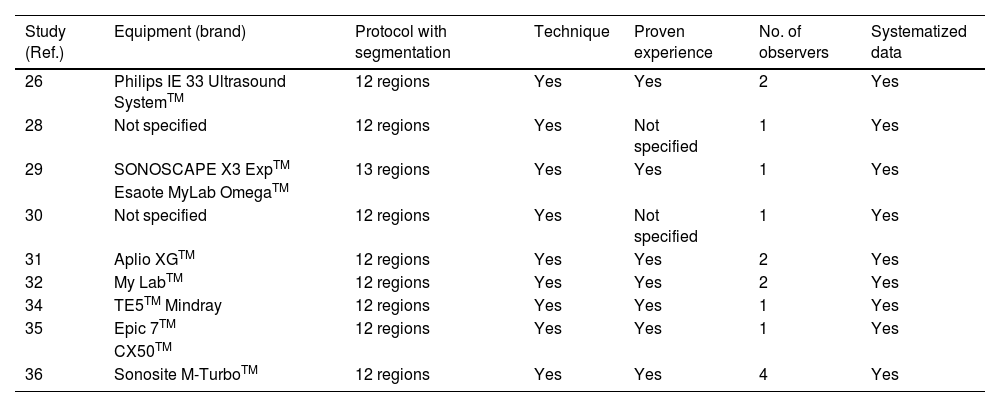
Assessment of the risk of bias was performed using the JBI Critical Appraisal Tools for observational studies. A negative response in any of the questionnaire sections would indicate that study quality was affected. All the studies included had a positive evaluation. In addition, the risk of observer-dependent biases for thoracic ultrasound data collection was analyzed. The following data were collected for each study: equipment (brand), whether the protocol for performing the technique was specified (segmentation of thoracic regions), the technique used, whether the observer had accredited experience, whether there was more than one observer, and whether the collection of ultrasound data was systematized. The results are shown in Table 2.

Table 2.

Quality of the ultrasound technique and operator observation.

Study (Ref.)  Equipment (brand)  Protocol with segmentation  Technique  Proven experience  No. of observers  Systematized data 
26  Philips IE 33 Ultrasound SystemTM  12 regions  Yes  Yes  Yes 
28  Not specified  12 regions  Yes  Not specified  Yes 
29SONOSCAPE X3 ExpTM  13 regionsYesYes1Yes
Esaote MyLab OmegaTM 
30  Not specified  12 regions  Yes  Not specified  Yes 
31  Aplio XGTM  12 regions  Yes  Yes  Yes 
32  My LabTM  12 regions  Yes  Yes  Yes 
34  TE5TM Mindray  12 regions  Yes  Yes  Yes 
35Epic 7TM  12 regionsYesYes1Yes
CX50TM 
36  Sonosite M-TurboTM  12 regions  Yes  Yes  Yes 
Discussion

Lung ultrasound abnormalities between two and six months after COVID-19 are frequent and their intensity is proportional to the severity of the initial disease.

The ultrasound abnormalities most frequently found in the studies were pleural line irregularities, B-lines, and subpleural consolidations. The frequency of pleural line irregularities was between 52% and 94% at 60 days28,32 and 22.5% at four months.28 The frequency of B-lines was between 40% and 70% at 60 days,28,31 between 30% and 60% at 90 days,26,29 and 20% and 30% at 120 days.28,30 Pleural line irregularities and B-lines were the most frequent findings and can translate into the presence of interstitial lung occupation in the periphery of the lung, which is compatible in this context and in the absence of other causes with a pulmonary syndrome. The frequency of subpleural consolidations was between 15% and 45% at 60 days,28,31 32% at 120 days,28 and 10% at 150 days.31

In the analysis by thoracic segments, a predominance of ultrasound abnormalities in the basal and posterior regions of the thorax has been described.28,29,35,36

The greater the severity of the initial disease, the higher the prevalence of lung ultrasound abnormalities in follow-up. As has been discussed, in Alharthy’s study,28 there are clearly more ultrasound findings in non-survivors versus survivors during acute infection (B-lines in 93.2% vs. 61%, pleural line irregularities in 90% vs. 70%, and consolidations in 77% vs. 51%). The study by Hernández-Píriz29 performed a subgroup analysis according to ARDS severity and found a higher frequency of ultrasound abnormalities in the most severe patients. The Fortini study30 only included patients with non-severe disease and noted mild lung ultrasound findings at follow-up. In a study at 180 days,35 the frequency of ultrasound abnormalities was higher than in other studies with a shorter time period. This could be explained by a greater severity of initial disease in these patients. A study that did not include any patients that had severe acute infection had 1% condensations at 90 days.29 Studies are needed to evaluate, according to the severity of the infection, the pulmonary ultrasound findings and the possible influencing factors, such as the severity of the initial infection, involvement of inflammatory mediators, the need for ventilatory support, and more.

The frequency of ultrasound abnormalities decreases over time. In the study by Hernández-Píriz,29 the frequency of abnormalities was 93.8% in the lung base at hospital admission and 30.9% in the same region at 90 days. In regard to pleural line irregularities, there was a decrease from 52% to 22% in the period from 60 to 120 days.28 Regarding B-lines, a decrease from 40% to 20% at two months was described.28 For subpleural consolidations, a slower decrease over time was observed, with some studies describing a reduction from 44% to 32% between the second and fourth month28 and from 69% to 57% between the second and fifth months.31 Subpleural consolidations occur in cases of greater infection involvement and therefore, the slow resolution could be related to this greater severity.

Studies have analyzed the correlation between lung ultrasound and CT scan findings and determined a high concordance.38,39 Haurylenka’s study38 analyzed 39 patients who had a CT scan immediately followed by a 12-region chest ultrasound and found a ROC curve for the ultrasound diagnosis of pneumonia of 0.97. In a study of 39 patients on ICU admission, a strong correlation was observed between thoracic ultrasound and CT scan findings, with an ROC curve of 0.953.39 This concordance remains high when pulmonary signs were analyzed after hospital discharge.28,36 A study of 352 patients between two and five months after pneumonia found a sensitivity of 94.2% and a negative predictive value of 94.7% for ultrasound.36 These results support the claim that lung ultrasound monitoring of patients is safe and does not subject patients to radiation, in addition to the technical and financial advantages of the test.

Limitations

Ultrasound is an operator-dependent imaging modality and can also be influenced by the patient’s physical characteristics and collaboration. There are different protocols for performing the ultrasound technique that segment the thorax into six,21,22 eight,40 12,22 or 14 regions.23,41 In the studies analyzed, the 12-region protocol was preferentially used. Although the studies analyzed collected ultrasound data in a systematic way, it was not uniform. Systematic data collection according to thoracic segmentation models benefits comparison among studies.

Under normal conditions, the anatomical characteristics themselves limit ultrasound lung examination due to the bone structures and the mediastinum. B-lines can be present in healthy individuals due to the gravitational component that increases hydrostatic pressure in the bases. These considerations must be taken into account in the interpretation and dissemination of the findings observed.

To date, the correlation of lung ultrasound findings with quality of life and persistence of symptoms has not been analyzed in depth. More studies are needed to evaluate lung ultrasound abnormalities in follow-up after hospital discharge; in patients who have had different degrees of lung involvement; and to establish the clinical, functional, and chest CT correlation.

Conclusions

The frequency of lung ultrasound manifestations decreases over time with a speed related to the severity of the initial infection. There seems to be good concordance between ultrasound and chest CT scan findings, but more studies are needed to support this observation and also to correlate lung ultrasound findings with severity of the initial infection, quality of life, laboratory parameters, and respiratory function tests.

Funding

The authors declare that they have received no financial compensation in relation to this article and have not received any funding.

Acknowledgments

To Dr. Francisco Martos Pérez and Dr. Cristina Asencio Méndez for reviewing the manuscript and special thanks to Dr. Javier García Alegría for his collaboration.

References
[1]
W. Guan, Z. Ni, Y. Hu, W. Liang, C. Ou, J. He, et al.
Clinical characteristics of coronavirus disease 2019 in China.
N Engl J Med., 382 (2009), pp. 1708-1720
[2]
WHO. (n.d.) (2020) WHO Director-General’s opening remarks at the media briefing on COVID-19—11. https://www.who.int/dg/speeches/detail/who-director-general-s-opening-remarks-at-themedia-brief​ing-on-covid-19 Accessed 11 Mar 2020.
[3]
WHO. (n.d.) (2021). COVID-19 Weekly Epidemiological Update. https://www.who.int/emergencies/diseases/novel-coronavirus-2019/situation-reports Accessed 14 May 2023.
[4]
X. Yang, Y. Yu, J. Xu, H. Shu, J. Xia, H. Liu, et al.
Clinical course and outcomes of critically ill patients with SARS-CoV-2 pneumonia in Wuhan, China: a single-centered, retrospective, observational study.
Lancet Respir Med., 8 (2020), pp. 475-481
[5]
G. Grasselli, A. Zangrillo, A. Zanella, M. Antonelli, L. Cabrini, A. Castelli, et al.
Baseline characteristics and outcomes of 1591 patients infected with SARS-CoV-2 admitted to ICUs of the Lombardy Region, Italy.
JAMA., 323 (2020), pp. 1574-1581
[6]
P. Metha, D.F. McAuley, M. Brown, E. Sanchez, R.S. Tattersall, J. Manson.
COVID-19: consider cytokine storm syndromes and immunosuppression.
Lancet., 395 (2020), pp. 1034-1035
[7]
D. Groff, A. Sun, A.E. Ssentongo, M.B. Djibril, N. Parsons, R.P. Govinda, et al.
Short-term and long-term rates os postacute sequelae of SARS-CoV-2 infection: a systematic review.
[8]
P.M. George, S.L. Barratt, R. Condliffe, S.J. Desai, A. Devaraj, I. Forrest, et al.
Respiratory follow-up of patients with COVID-19 pneumonia.
Thorax., 75 (2020), pp. 1009-1016
[9]
O. Sibila, M. Molina-Molina, C. Valenzuela, A. Ríos-Cortés, A. Arbillaga-Etxarri, Y. Torralba, et al.
Documento de consenso de la Sociedad Española de Neumología y Cirugía Torácica (SEPAR) para el seguimiento clínico post-COVID-19.
Open Respir Arch., 2 (2020), pp. 278-283
[10]
H. Wong, H. Lam, A. Fong, S. Leung, T. Chin, C. Lo, et al.
Frequency and distribution of chest radiographics findings in patients positive for COVID-19.
Radiology., 296 (2020), pp. 72-78
[11]
Z. Zu, M. Jiang, P. Xu, W. Chen, Q. Ni, G. Lu, et al.
Coronavirus disease 2019 (COVID-19): a perspective from China.
Radiology., 296 (2020), pp. 15-25
[12]
T. Ai, Z. Yang, H. Hou, C. Zhan, C. Chen, W. Lv, et al.
Correlation of chest and RT-PCR testing for coronavirus disease 2019 (COVID-19) in China: a report of 1014 cases.
Radiology., 296 (2020), pp. 32-40
[13]
H. Kim, H. Hong, S. Yoon.
Diagnostic performance of CT and reverse transcriptase polymerase chain reaction for coronavirus disease 2019: a meta-analysis.
Radiology., 296 (2020), pp. 145-155
[14]
J. Kanne, B. Little, J. Chung, B. Elicker, L. Ketai.
Essentials for radiologists on COVID-19: an uptdate-radiology scientific expert panel.
Radiology., 296 (2020), pp. 113-114
[15]
F. Pan, T. Ye, P. Sun, S. Gui, B. Liang, L. Li, et al.
Time course of lung changes at chest CT during recovery from coronavirus disease 2019 (COVID-19).
Radiology., 295 (2020), pp. 715-721
[16]
J. Solomon, B. Heyman, J.P. Ko, R. Condos, D.A. Lynch.
CT of post-acute lung complications of COVID-19.
Radiology., 301 (2021), pp. 383-395
[17]
X. Han, Y. Fan, O. Alwalid, N. Li, X. Jia, M. Yuan, et al.
Six-month follow-up chest CT findings after severe COVID-19 pneumonia.
Radiology., 299 (2021), pp. 177-186
[18]
D. Lichtestein, O. Axler.
Intensive use of general ultrasound in the intensive care unit. Prospective study of 150 consecutive patients.
Intensive Care Med., 19 (1993), pp. 353-355
[19]
C. Sorlini, M. Femia, G. Nattino, P. Bellone, E. Gesu, P. Francione, et al.
The role of lung ultrasound as a frontline diagnostic tool in the era of COVID-19 outbreak.
Intern Emerg Med., 16 (2020), pp. 749-756
[20]
G. Lepri, M. Orlandi, C. Lazzeri, C. Bruni, M. Hughes, M. Bonizzoli, et al.
The emerging role of lung ultrasound in COVID-19 pneumonia.
Eur J Rheumatol., 7 (2020), pp. 5129-5133
[21]
M.J. Smith, S.A. Hayward, S.M. Innes, A.S.C. Miller.
Point-of-care lung ultrasound in patients with COVID-19 – a narrative review.
Anaesthesia., 75 (2020), pp. 1096-1104
[22]
Q. Peng, X.T. Wang, L.N. Zhang, and Chinese Care Ultrasound Study Group (CCUSG).
Findings of lung ultrasonography of novel corona virus pneumonia during the 2019-2020 epidemic.
Intensive Care Med., 46 (2020), pp. 849-850
[23]
S. Sofia, A. Boccatonda, M. Montanari, M. Spampinato, D. D’ardes, G. Cocco, et al.
Thoracic ultrasound and SARS-COVID-19: a pictorial essay.
J Ultrasound., 23 (2020), pp. 217-221
[24]
M.J. Page, J.E. McKenzie, P.M. Bossuyt, I. Boutron, T.C. Hoffmann, C.D. Mulrow, et al.
The PRISMA 2020 statement: an updated guideline for reporting systematic reviews.
BMJ., 372 (2021), pp. 71
[25]
Z. Munn, T.H. Barker, S. Moola, C. Tufanaru, C. Stern, A. McArthur, et al.
Methodological quality of case series studies: an introduction to the JBI critical appraisal tool.
JBI Evidence Synthesis., 18 (2020), pp. 2127-2133
[26]
G. Giovannetti, L. Michele, M. Ceglie, P. Pierucci, A. Mirabile, M. Vita, et al.
Lung ultrasonography for long-term follow-up of COVID-19 survivors compared to chest CT scan.
[27]
A. Osman, R. Hashim.
Diaphragmatic and lung ultrasound application as new predictive indices for the weaning process in ICU patients.
Egypt J Radiol Nucl Med., 48 (2021), pp. 61-66
[28]
A. Alharthy, M. Abuhamdah, A. Balhamar, F. Faqihi, N. Nasim, S. Ahmad, et al.
Residual lung injury in patients recovering from COVID-19 critical illines: a prospective longitudinal Point-of-Care lung ultrasound study.
J Ultrasound Med., 40 (2020), pp. 1823-1838
[29]
A. Hernández-Píriz, Y. Tung-Chen, D. Jiménez-Virumbrales, I. Ayala-Larrañaga, R. Barba-Martín, J. Canora-Lebrato, et al.
Importance of lung ultrasound follow-up in patients who had recovered from coronavirus disease 2019: results from a prospective study.
[30]
A. Fortini, A. Torrigiane, S. Sbaragli, A. Lo Forte, A. Crociani, P. Cecchini, et al.
COVID-19: persistence of symptoms and lung alterations after 3-6 months from hospital discharge.
Infection., 49 (2021), pp. 1007-1015
[31]
A.J. Lopes, P.F. Litrento, B.C. Provenzano, A.S. Carneiro, L.B. Monneat, M.S. da Cal, et al.
Small airway dysfunction on impulse oscillometry and pathological signs on lung ultrasound are frequent in post-COVID-19 patients with persistent respiratory symptoms.
[32]
M. Ferioli, I. Prediletto, S. Bensai, S. Betti, F. Danielle, V. Di Scioscio, et al.
Spontaneus evolution of COVID-19 lung sequelae: results from a double step follow-up.
Respiration., 101 (2022), pp. 381-393
[33]
G. Volpicelli, M. Elbarbary, M. Blaivas, D.A. Lichtenstein, G. Mathis, A.W. Kirkpatrick, et al.
International Liaison Committee on Lung Ultrasound (ILC-LUS) for International Consensus Conference on Lung Ultrasound (ICC-LUS). International evidence-based recommendations for point-of-care lung ultrasound.
Intensive Care Med., 38 (2012), pp. 577-591
[34]
N. Gurbani, M. Acosta-Sorensen, D. Díaz-Pérez, J.M. Figueira-Goncalves, Y. Ramallo-Fariña, J.L. Trujillo-Castilla.
Clinical outcomes and lung ultrasound findings in COVID-19 follow up: calm comes after the storm?.
[35]
G. Russo, N. Flor, F. Casella, S. Ippolito, F. Leidi, G. Casazza, et al.
Lung ultrasound in the follow-up of severe COVID-19 pneumoniae: six months evaluation and comparison with CT.
[36]
D. Clofent, E. Polverino, A. Felipe, G. Granados, M. Arjona-Peris, J. Andreu, et al.
Lung ultrasound as a First-Line Test in the evaluation of post-COVID-19 pulmonary sequelae.
[37]
J.H. Warrick, M. Bhalla, S.I. Schabel, R.M. Silver.
High resolution computed tomography in early scleroderma lung disease.
J Rheumatol., 18 (1991), pp. 1520-1528
[38]
D. Haurylenka, V. Damantsevich, A. Filustsin, A. Damantsevich.
Diagnostic value of 12-zone lung ultrasound protocol for diagnosing COVID-19-associated pneumonia in outpatients.
[39]
D. Orlandi, D. Battaglini, C. Robba, M. Viganò, G. Bergamaschi, T. Mignatiti, et al.
Coronavirus disease 2019 phenotypes, lung ultrasound, chest computed tomography and clinical features in critically ill mechanically ventiled patients.
Ultrasound Med Biol., 12 (2021), pp. 3323-3332
[40]
S. Fox, S. Dugar.
Point-Of-Care ultrasound and COVID-19.
[41]
G. Soldati, A. Smargiassi, R. Inchingolo, D. Buonsenso, T. Perrone, D.F. Briganti, et al.
Proposal for International Standardization of the use of lung ultrasound for patients with COVID-19.
J Ultrasound Med., 39 (2020), pp. 1413-1419
Copyright © 2024. Elsevier España, S.L.U. and Sociedad Española de Medicina Interna (SEMI)
Download PDF
Idiomas
Revista Clínica Española (English Edition)
Article options
Tools
es en

¿Es usted profesional sanitario apto para prescribir o dispensar medicamentos?

Are you a health professional able to prescribe or dispense drugs?