Compartir
array:24 [
"pii" => "S0014256516301916"
"issn" => "00142565"
"doi" => "10.1016/j.rce.2016.11.004"
"estado" => "S300"
"fechaPublicacion" => "2017-04-01"
"aid" => "1333"
"copyright" => "Elsevier España, S.L.U. and Sociedad Española de Medicina Interna (SEMI)"
"copyrightAnyo" => "2016"
"documento" => "article"
"crossmark" => 1
"subdocumento" => "sco"
"cita" => "Rev Clin Esp. 2017;217:144-8"
"abierto" => array:3 [
"ES" => false
"ES2" => false
"LATM" => false
]
"gratuito" => false
"lecturas" => array:2 [
"total" => 41
"formatos" => array:2 [
"HTML" => 23
"PDF" => 18
]
]
"Traduccion" => array:1 [
"en" => array:19 [
"pii" => "S2254887417300188"
"issn" => "22548874"
"doi" => "10.1016/j.rceng.2016.11.002"
"estado" => "S300"
"fechaPublicacion" => "2017-04-01"
"aid" => "1333"
"copyright" => "Elsevier España, S.L.U. and Sociedad Española de Medicina Interna (SEMI)"
"documento" => "article"
"crossmark" => 1
"subdocumento" => "sco"
"cita" => "Rev Clin Esp. 2017;217:144-8"
"abierto" => array:3 [
"ES" => false
"ES2" => false
"LATM" => false
]
"gratuito" => false
"lecturas" => array:2 [
"total" => 5
"formatos" => array:2 [
"HTML" => 2
"PDF" => 3
]
]
"en" => array:12 [
"idiomaDefecto" => true
"cabecera" => "<span class="elsevierStyleTextfn">Brief Original</span>"
"titulo" => "Examination of cytological smears and cell blocks of pleural fluid: Complementary diagnostic value for malignant effusions"
"tienePdf" => "en"
"tieneTextoCompleto" => "en"
"tieneResumen" => array:2 [
0 => "en"
1 => "es"
]
"paginas" => array:1 [
0 => array:2 [
"paginaInicial" => "144"
"paginaFinal" => "148"
]
]
"titulosAlternativos" => array:1 [
"es" => array:1 [
"titulo" => "Examen del frotis citológico y bloque celular del líquido pleural: valor diagnóstico complementario en los derrames malignos"
]
]
"contieneResumen" => array:2 [
"en" => true
"es" => true
]
"contieneTextoCompleto" => array:1 [
"en" => true
]
"contienePdf" => array:1 [
"en" => true
]
"autores" => array:1 [
0 => array:2 [
"autoresLista" => "J.M. Porcel, M. Quirós, S. Gatius, S. Bielsa"
"autores" => array:4 [
0 => array:2 [
"nombre" => "J.M."
"apellidos" => "Porcel"
]
1 => array:2 [
"nombre" => "M."
"apellidos" => "Quirós"
]
2 => array:2 [
"nombre" => "S."
"apellidos" => "Gatius"
]
3 => array:2 [
"nombre" => "S."
"apellidos" => "Bielsa"
]
]
]
]
]
"idiomaDefecto" => "en"
"Traduccion" => array:1 [
"en" => array:9 [
"pii" => "S0014256516301916"
"doi" => "10.1016/j.rce.2016.11.004"
"estado" => "S300"
"subdocumento" => ""
"abierto" => array:3 [
"ES" => false
"ES2" => false
"LATM" => false
]
"gratuito" => false
"lecturas" => array:1 [
"total" => 0
]
"idiomaDefecto" => "en"
"EPUB" => "https://multimedia.elsevier.es/PublicationsMultimediaV1/item/epub/S0014256516301916?idApp=WRCEE"
]
]
"EPUB" => "https://multimedia.elsevier.es/PublicationsMultimediaV1/item/epub/S2254887417300188?idApp=WRCEE"
"url" => "/22548874/0000021700000003/v1_201703290108/S2254887417300188/v1_201703290108/en/main.assets"
]
]
"itemSiguiente" => array:18 [
"pii" => "S0014256517300279"
"issn" => "00142565"
"doi" => "10.1016/j.rce.2017.01.003"
"estado" => "S300"
"fechaPublicacion" => "2017-04-01"
"aid" => "1354"
"copyright" => "Elsevier España, S.L.U. and Sociedad Española de Medicina Interna (SEMI)"
"documento" => "simple-article"
"crossmark" => 1
"subdocumento" => "edi"
"cita" => "Rev Clin Esp. 2017;217:149-50"
"abierto" => array:3 [
"ES" => false
"ES2" => false
"LATM" => false
]
"gratuito" => false
"lecturas" => array:2 [
"total" => 107
"formatos" => array:2 [
"HTML" => 61
"PDF" => 46
]
]
"es" => array:10 [
"idiomaDefecto" => true
"cabecera" => "<span class="elsevierStyleTextfn">Editorial</span>"
"titulo" => "Utilidad diagnóstica del recuento leucocitario total y diferencial del líquido pleural"
"tienePdf" => "es"
"tieneTextoCompleto" => "es"
"paginas" => array:1 [
0 => array:2 [
"paginaInicial" => "149"
"paginaFinal" => "150"
]
]
"titulosAlternativos" => array:1 [
"en" => array:1 [
"titulo" => "Diagnostic usefulness of pleural fluid total leukocyte count and differential"
]
]
"contieneTextoCompleto" => array:1 [
"es" => true
]
"contienePdf" => array:1 [
"es" => true
]
"autores" => array:1 [
0 => array:2 [
"autoresLista" => "J.M. Porcel"
"autores" => array:1 [
0 => array:2 [
"nombre" => "J.M."
"apellidos" => "Porcel"
]
]
]
]
]
"idiomaDefecto" => "es"
"EPUB" => "https://multimedia.elsevier.es/PublicationsMultimediaV1/item/epub/S0014256517300279?idApp=WRCEE"
"url" => "/00142565/0000021700000003/v1_201703290111/S0014256517300279/v1_201703290111/es/main.assets"
]
"itemAnterior" => array:19 [
"pii" => "S0014256517300280"
"issn" => "00142565"
"doi" => "10.1016/j.rce.2016.12.014"
"estado" => "S300"
"fechaPublicacion" => "2017-04-01"
"aid" => "1355"
"copyright" => "Elsevier España, S.L.U. and Sociedad Española de Medicina Interna (SEMI)"
"documento" => "article"
"crossmark" => 1
"subdocumento" => "fla"
"cita" => "Rev Clin Esp. 2017;217:136-43"
"abierto" => array:3 [
"ES" => false
"ES2" => false
"LATM" => false
]
"gratuito" => false
"lecturas" => array:2 [
"total" => 79
"formatos" => array:2 [
"HTML" => 43
"PDF" => 36
]
]
"es" => array:13 [
"idiomaDefecto" => true
"cabecera" => "<span class="elsevierStyleTextfn">ORIGINAL</span>"
"titulo" => "Comportamiento de las células nucleadas en los distintos tipos de derrame pleural"
"tienePdf" => "es"
"tieneTextoCompleto" => "es"
"tieneResumen" => array:2 [
0 => "es"
1 => "en"
]
"paginas" => array:1 [
0 => array:2 [
"paginaInicial" => "136"
"paginaFinal" => "143"
]
]
"titulosAlternativos" => array:1 [
"en" => array:1 [
"titulo" => "Behaviour of nucleated cells in various types of pleural effusion"
]
]
"contieneResumen" => array:2 [
"es" => true
"en" => true
]
"contieneTextoCompleto" => array:1 [
"es" => true
]
"contienePdf" => array:1 [
"es" => true
]
"resumenGrafico" => array:2 [
"original" => 0
"multimedia" => array:7 [
"identificador" => "fig0005"
"etiqueta" => "Figura 1"
"tipo" => "MULTIMEDIAFIGURA"
"mostrarFloat" => true
"mostrarDisplay" => false
"figura" => array:1 [
0 => array:4 [
"imagen" => "gr1.jpeg"
"Alto" => 1721
"Ancho" => 1667
"Tamanyo" => 374748
]
]
"descripcion" => array:1 [
"es" => "<p id="spar0045" class="elsevierStyleSimplePara elsevierViewall">Valores de los porcentajes de neutrófilos (Neu.) y linfocitos (Linf.) de cada paciente, y su mediana, según la etilogía del derrame pleural.</p> <p id="spar0050" class="elsevierStyleSimplePara elsevierViewall">DPP: derrame pleural paraneumónico; IC: insuficiencia cardiaca; NEO: neoplasia; OE: otros exudados; OT: otros trasudados; TB: tuberculosis.</p>"
]
]
]
"autores" => array:1 [
0 => array:2 [
"autoresLista" => "L. Ferreiro, T. Pereiro, E. San José, M.E. Toubes, J. Suárez-Antelo, J.M. Álvarez Dobaño, F.J. González Barcala, N. Rodríguez Núñez, A. Lama, L. Valdés"
"autores" => array:10 [
0 => array:2 [
"nombre" => "L."
"apellidos" => "Ferreiro"
]
1 => array:2 [
"nombre" => "T."
"apellidos" => "Pereiro"
]
2 => array:2 [
"nombre" => "E."
"apellidos" => "San José"
]
3 => array:2 [
"nombre" => "M.E."
"apellidos" => "Toubes"
]
4 => array:2 [
"nombre" => "J."
"apellidos" => "Suárez-Antelo"
]
5 => array:2 [
"nombre" => "J.M."
"apellidos" => "Álvarez Dobaño"
]
6 => array:2 [
"nombre" => "F.J."
"apellidos" => "González Barcala"
]
7 => array:2 [
"nombre" => "N."
"apellidos" => "Rodríguez Núñez"
]
8 => array:2 [
"nombre" => "A."
"apellidos" => "Lama"
]
9 => array:2 [
"nombre" => "L."
"apellidos" => "Valdés"
]
]
]
]
]
"idiomaDefecto" => "es"
"Traduccion" => array:1 [
"en" => array:9 [
"pii" => "S2254887417300097"
"doi" => "10.1016/j.rceng.2016.12.006"
"estado" => "S300"
"subdocumento" => ""
"abierto" => array:3 [
"ES" => false
"ES2" => false
"LATM" => false
]
"gratuito" => false
"lecturas" => array:1 [
"total" => 0
]
"idiomaDefecto" => "en"
"EPUB" => "https://multimedia.elsevier.es/PublicationsMultimediaV1/item/epub/S2254887417300097?idApp=WRCEE"
]
]
"EPUB" => "https://multimedia.elsevier.es/PublicationsMultimediaV1/item/epub/S0014256517300280?idApp=WRCEE"
"url" => "/00142565/0000021700000003/v1_201703290111/S0014256517300280/v1_201703290111/es/main.assets"
]
"en" => array:18 [
"idiomaDefecto" => true
"cabecera" => "<span class="elsevierStyleTextfn">Brief Original</span>"
"titulo" => "Examination of cytological smears and cell blocks of pleural fluid: Complementary diagnostic value for malignant effusions"
"tieneTextoCompleto" => true
"paginas" => array:1 [
0 => array:2 [
"paginaInicial" => "144"
"paginaFinal" => "148"
]
]
"autores" => array:1 [
0 => array:4 [
"autoresLista" => "J.M. Porcel, M. Quirós, S. Gatius, S. Bielsa"
"autores" => array:4 [
0 => array:4 [
"nombre" => "J.M."
"apellidos" => "Porcel"
"email" => array:1 [
0 => "jporcelp@yahoo.es"
]
"referencia" => array:2 [
0 => array:2 [
"etiqueta" => "<span class="elsevierStyleSup">a</span>"
"identificador" => "aff0005"
]
1 => array:2 [
"etiqueta" => "<span class="elsevierStyleSup">*</span>"
"identificador" => "cor0005"
]
]
]
1 => array:3 [
"nombre" => "M."
"apellidos" => "Quirós"
"referencia" => array:1 [
0 => array:2 [
"etiqueta" => "<span class="elsevierStyleSup">a</span>"
"identificador" => "aff0005"
]
]
]
2 => array:3 [
"nombre" => "S."
"apellidos" => "Gatius"
"referencia" => array:1 [
0 => array:2 [
"etiqueta" => "<span class="elsevierStyleSup">b</span>"
"identificador" => "aff0010"
]
]
]
3 => array:3 [
"nombre" => "S."
"apellidos" => "Bielsa"
"referencia" => array:1 [
0 => array:2 [
"etiqueta" => "<span class="elsevierStyleSup">a</span>"
"identificador" => "aff0005"
]
]
]
]
"afiliaciones" => array:2 [
0 => array:3 [
"entidad" => "Pleural Medicine Unit, Arnau de Vilanova University Hospital, Lleida, Spain"
"etiqueta" => "a"
"identificador" => "aff0005"
]
1 => array:3 [
"entidad" => "Department of Pathology, Arnau de Vilanova University Hospital, Lleida, Spain"
"etiqueta" => "b"
"identificador" => "aff0010"
]
]
"correspondencia" => array:1 [
0 => array:3 [
"identificador" => "cor0005"
"etiqueta" => "⁎"
"correspondencia" => "Corresponding author."
]
]
]
]
"titulosAlternativos" => array:1 [
"es" => array:1 [
"titulo" => "Examen del frotis citológico y bloque celular del líquido pleural: valor diagnóstico complementario en los derrames malignos"
]
]
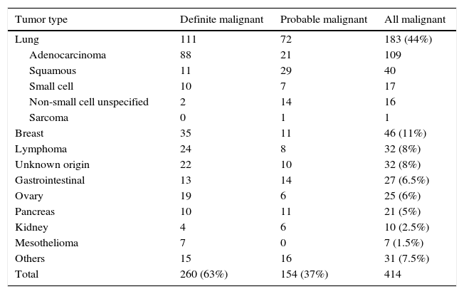
"textoCompleto" => "<span class="elsevierStyleSections"><span id="sec0005" class="elsevierStyleSection elsevierViewall"><span class="elsevierStyleSectionTitle" id="sect0065">Introduction</span><p id="par0005" class="elsevierStylePara elsevierViewall">Cytologic examination of pleural fluid is the easiest and least invasive way to diagnose malignant effusions, although it has limited sensitivity.<a class="elsevierStyleCrossRef" href="#bib0090"><span class="elsevierStyleSup">1</span></a> The routine cytopreparatory technique consists of sample centrifugation, smearing of the cell deposit and Papanicolau staining.<a class="elsevierStyleCrossRef" href="#bib0095"><span class="elsevierStyleSup">2</span></a> Scientific guidelines indicate that preparing cell blocks (CBs) from pleural effusion samples, in addition to smears, to allow for “microhistology” of the cellular solid portion may lead to greater diagnostic accuracy.<a class="elsevierStyleCrossRef" href="#bib0100"><span class="elsevierStyleSup">3</span></a> However, the magnitude of this diagnostic yield increase has barely been reported. Moreover, since CB methods can be time consuming and labor intensive they may not be routine in some centers, only being performed at the discretion of the pathologist or clinician. Our aim was to analyze, in the largest series reported to date, the usefulness of CBs beyond conventional cytological smears in the diagnosis of malignant effusions. Biochemical fluid characteristics predictive of the generation of inappropriate or insufficient CB material were also addressed.</p></span><span id="sec0010" class="elsevierStyleSection elsevierViewall"><span class="elsevierStyleSectionTitle" id="sect0070">Methods</span><p id="par0010" class="elsevierStylePara elsevierViewall">The medical records of all consecutive patients at our institution between October 2010 and June 2016 whose pleural fluids had been sent for conventional cytology and/or CB examination, based on the attending physician's criterion, were retrospectively reviewed. The local ethics committee approved the study protocol.</p><p id="par0015" class="elsevierStylePara elsevierViewall">Pleural fluid specimens were collected in heparinized tubes. For routine cytology, 5<span class="elsevierStyleHsp" style=""></span>mL of fluid were centrifuged at 2000<span class="elsevierStyleHsp" style=""></span>rpm for 10<span class="elsevierStyleHsp" style=""></span>min, the supernatant discarded, and the glass slides prepared and stained with Papanicolaou stain. For CB preparation, 10<span class="elsevierStyleHsp" style=""></span>mL of material were centrifuged as above, the supernatant decanted, and the cell button obtained (with the addition of molten agar if necessary) fixed in 10% formaldehyde, paraffin embedded and stained with hematoxylin–eosin. Immunocytochemistry panels on CB sections were applied according to the suspected tumor type and cytomorphology.</p><p id="par0020" class="elsevierStylePara elsevierViewall">An effusion was categorized as definite malignant if malignant cells were detected upon cytological examination of pleural fluid or biopsy specimens. A diagnosis of probable malignant effusion was made on patients who met the following: (a) a known primary tumor or extrapleural metastases of undetermined origin, and (b) a pleural exudate with negative fluid cytological findings, after ruling out other potential causes of fluid accumulation by clinical data (e.g. negative pleural fluid cultures, low pleural adenosine deaminase levels), imaging (e.g. CT angiography) and, in selected cases, pleural biopsy. All patients with probable MPE were followed up long enough to determine whether alternative causes of the effusion became clinically apparent.</p><p id="par0025" class="elsevierStylePara elsevierViewall">Sensitivity of fluid smears and CBs were calculated. The Kruskal–Wallis test was used to compare pleural fluid biochemistries (i.e., red and white blood cell counts, differential white cell count, protein, lactate dehydrogenase (LDH), glucose, adenosine deaminase, pH and C-reactive protein) between patients with representative and suboptimal CB material. The statistical significance level was set at 0.05 (two-tailed).</p></span><span id="sec0015" class="elsevierStyleSection elsevierViewall"><span class="elsevierStyleSectionTitle" id="sect0075">Results</span><p id="par0030" class="elsevierStylePara elsevierViewall">During the study period, 632 cytological smears and 554 CBs from 414 patients (median age 70 years, 56% men) who had malignant effusions were examined. The etiological distribution of the primary tumors is displayed in <a class="elsevierStyleCrossRef" href="#tbl0005">Table 1</a>. Also, there were 726 patients eventually diagnosed with a benign pleural condition for whom pleural fluid cytological analyses were ordered (a total of 945 smears and 507 CBs) and yielded negative results; all were excluded for the purpose of the study.</p><elsevierMultimedia ident="tbl0005"></elsevierMultimedia><p id="par0035" class="elsevierStylePara elsevierViewall">Although all effusions were submitted to a first cytologic examination, CBs were initially obtained from 380 (92%) specimens, but 46 (12%) yielded suboptimal material. Whether a first cytological smear or CB examination was used, both had identical sensitivity for identifying malignancy: 44% (95% CI 39–50%). Notably, of 184 cancer patients who had a first negative cytological exam, CB was diagnostic of malignancy in 18 (10%, 95% CI 6–15%). Conversely, among the 186 patients with a first negative CB examination, cytological smears were positive in 20 (11%, 95% CI 7–16%). If any further cytology and CB assessments in patients with previous negative results were considered, the global discrepancy was as follows: 11% (95% CI 7–17%) smears negative/CBs positive, and 15% (95% CI 11–21%) smears positive/CBs negative. Overall, 74 of the 554 CB preparations (13%, 95% CI 11–16%) resulted in inappropriate material for interpretation or diagnosis.</p><p id="par0040" class="elsevierStylePara elsevierViewall">Immunocytochemical studies were performed on 157 (34%) CBs, particularly in those regarded as positive by cytomorphology (85% vs 1.7%), with a median of 3 (25th to 75th percentiles 1–4) immunostains per case. From a total of 480 immunostainings, the most commonly used, in order of decreasing frequency, were TTF-1 (138, 29%), estrogen receptor (60, 13%), CK 7 (47, 47%), CK 20 (43, 9%), p63 (30, 6%), calretinin (25, 5%), mammaglobin (21, 4%), WT1 (17, 4%), and CK 19 (15, 3%), among others.</p><p id="par0045" class="elsevierStylePara elsevierViewall">The sequential sensitivity of separated pleural fluid smears, CBs or their combination for establishing the diagnosis of malignant pleural effusions is shown in <a class="elsevierStyleCrossRef" href="#tbl0010">Table 2</a>. In general, cytological studies eventually identified 233 (56%, 95% CI 51–61%) malignant cases. It was also demonstrated that the yield by sending more than two specimens is negligible.</p><elsevierMultimedia ident="tbl0010"></elsevierMultimedia><p id="par0050" class="elsevierStylePara elsevierViewall">As compared with the reported positive and negative CBs, suboptimal CB materials exhibited significantly lower median red blood cell (10,250/μL vs 3600/μL vs 2200/μL, <span class="elsevierStyleItalic">p</span><span class="elsevierStyleHsp" style=""></span><<span class="elsevierStyleHsp" style=""></span>0.01) and leukocyte counts (905/μL vs 1158/μL vs 392/μL, <span class="elsevierStyleItalic">p</span><span class="elsevierStyleHsp" style=""></span><<span class="elsevierStyleHsp" style=""></span>0.01). Furthermore, higher median LDH levels (732<span class="elsevierStyleHsp" style=""></span>U/L vs 434<span class="elsevierStyleHsp" style=""></span>U/L vs 394<span class="elsevierStyleHsp" style=""></span>U/L, <span class="elsevierStyleItalic">p</span><span class="elsevierStyleHsp" style=""></span><<span class="elsevierStyleHsp" style=""></span>0.01) and decreased pH values (7.42 vs 7.46 vs 7.47, <span class="elsevierStyleItalic">p</span><span class="elsevierStyleHsp" style=""></span><<span class="elsevierStyleHsp" style=""></span>0.01) were associated with a greater likelihood of obtaining a positive CB result rather than a negative one or an insufficient CB.</p></span><span id="sec0020" class="elsevierStyleSection elsevierViewall"><span class="elsevierStyleSectionTitle" id="sect0080">Discussion</span><p id="par0055" class="elsevierStylePara elsevierViewall">This study suggests that the examination of CBs alongside fluid smears is a valuable first step in the diagnostic workup for any suspected malignant effusion. Conventional pleural fluid cytology is negative for malignant cells in up to 40% of malignant effusions, and in an even greater proportion in mesotheliomas and squamous cell lung carcinomas.<a class="elsevierStyleCrossRef" href="#bib0105"><span class="elsevierStyleSup">4</span></a> Although CB is an old methodology for the diagnosis of serous effusions, few studies have suggested its potential diagnostic superiority as compared to cytological smears.<a class="elsevierStyleCrossRefs" href="#bib0110"><span class="elsevierStyleSup">5–12</span></a> In brief, they have reported that CBs increase the diagnostic yield for malignancy by 5–15% over routine cytology. However, the inclusion of a small number of cases (no study recruited more than 60 malignant effusions) limits the validity of the results. Even an earlier report casts doubt on the cost-effectiveness of preparing CBs and advocated for cytology smears alone.<a class="elsevierStyleCrossRef" href="#bib0150"><span class="elsevierStyleSup">13</span></a> Of note, a recent survey among last two-year Spanish residents of Pneumology or Internal Medicine showed that only 16% of 139 responders in their clinical practice actively ordered a CB when confronted with a suspected malignant effusion, whereas the remaining either never did (27%) or left the decision to the pathologist discretion (57%).<a class="elsevierStyleCrossRef" href="#bib0155"><span class="elsevierStyleSup">14</span></a> Therefore, despite clinical guidelines which consider CB techniques to be a useful addition for diagnostic purposes,<a class="elsevierStyleCrossRefs" href="#bib0100"><span class="elsevierStyleSup">3,15</span></a> it appears that too little attention has been paid to this recommendation.</p><p id="par0060" class="elsevierStylePara elsevierViewall">CBs have some advantages over cytological smears which may facilitate the diagnosis of malignancy<a class="elsevierStyleCrossRef" href="#bib0165"><span class="elsevierStyleSup">16</span></a>: (a) a better appreciation of tissue architecture (e.g., acinar or papillary structures, intracytoplasmatic mucin), (b) an easier cytomorphological distinction between reactive mesothelial cells and mesothelioma or metastatic adenocarcinoma, and (c) the possibility of processing multiple sections for immunocytochemistry. Of interest, smears and CBs were found to have the same sensitivity (44% on a first specimen), but discrepant results (i.e., negative smears and positive CBs, or vice versa in 11% and 15% of cases, respectively) pointed to their complementary value. Therefore, CBs are not a substitute for fluid smears, but rather an additional technique for increasing diagnostic accuracy. It was also found that pleural fluids containing low red blood and/or leukocyte cell counts are likely more unsuitable for CB preparation.</p><p id="par0065" class="elsevierStylePara elsevierViewall">Our reported sensitivity for a first cytological pleural fluid specimen (44%) is on the lower end of the spectrum for malignant effusion diagnoses. However, it is in line with our previous series from the 1993–2006 period in which the yield of a first cytological analysis among 466 patients with malignant effusions was reported to be 48.5%.<a class="elsevierStyleCrossRef" href="#bib0170"><span class="elsevierStyleSup">17</span></a> It should be highlighted that we only send 5<span class="elsevierStyleHsp" style=""></span>mL of pleural fluid for smears preparation, and that the incidence of positive results depends on the volume of pleural fluid submitted (the larger the amount the greater the diagnostic accuracy)<a class="elsevierStyleCrossRef" href="#bib0090"><span class="elsevierStyleSup">1</span></a> and other factors such as the number of specimens evaluated (i.e., more than two specimens do not increase sensitivity sufficiently to be clinically meaningful), the way in which the specimens are examined (e.g., CBs along with smears, as this study supports), the tumor type, and the experience of the cytopathologist.</p><p id="par0070" class="elsevierStylePara elsevierViewall">This study has limitations. First, it is retrospective. Second, pathologists were not blinded of clinical data and the interpretation of CBs may also have been influenced by the results of cytology smears, which however, is a reflection of daily practice. Finally, in a third of pleural effusions the malignant nature was not confirmed by pleural biopsy, though it was based on solid clinical grounds.</p><p id="par0075" class="elsevierStylePara elsevierViewall">In conclusion, this study suggests that clinicians should be encouraged to request, along with the conventional routine cytology, CBs of pleural fluid on which immunocytochemical staining can be performed, in order to increase diagnostic sensitivity and establish a more definitive cytopathological diagnosis, whenever a malignant condition is suspected.</p></span><span id="sec0025" class="elsevierStyleSection elsevierViewall"><span class="elsevierStyleSectionTitle" id="sect0085">Conflict of interests</span><p id="par0080" class="elsevierStylePara elsevierViewall">The authors declare no conflict of interests.</p></span></span>"
"textoCompletoSecciones" => array:1 [
"secciones" => array:10 [
0 => array:3 [
"identificador" => "xres821255"
"titulo" => "Abstract"
"secciones" => array:4 [
0 => array:2 [
"identificador" => "abst0005"
"titulo" => "Objectives"
]
1 => array:2 [
"identificador" => "abst0010"
"titulo" => "Patients and methods"
]
2 => array:2 [
"identificador" => "abst0015"
"titulo" => "Results"
]
3 => array:2 [
"identificador" => "abst0020"
"titulo" => "Conclusions"
]
]
]
1 => array:2 [
"identificador" => "xpalclavsec818247"
"titulo" => "Keywords"
]
2 => array:3 [
"identificador" => "xres821256"
"titulo" => "Resumen"
"secciones" => array:4 [
0 => array:2 [
"identificador" => "abst0025"
"titulo" => "Objetivos"
]
1 => array:2 [
"identificador" => "abst0030"
"titulo" => "Pacientes y métodos"
]
2 => array:2 [
"identificador" => "abst0035"
"titulo" => "Resultados"
]
3 => array:2 [
"identificador" => "abst0040"
"titulo" => "Conclusiones"
]
]
]
3 => array:2 [
"identificador" => "xpalclavsec818246"
"titulo" => "Palabras clave"
]
4 => array:2 [
"identificador" => "sec0005"
"titulo" => "Introduction"
]
5 => array:2 [
"identificador" => "sec0010"
"titulo" => "Methods"
]
6 => array:2 [
"identificador" => "sec0015"
"titulo" => "Results"
]
7 => array:2 [
"identificador" => "sec0020"
"titulo" => "Discussion"
]
8 => array:2 [
"identificador" => "sec0025"
"titulo" => "Conflict of interests"
]
9 => array:1 [
"titulo" => "References"
]
]
]
"pdfFichero" => "main.pdf"
"tienePdf" => true
"fechaRecibido" => "2016-09-20"
"fechaAceptado" => "2016-11-09"
"PalabrasClave" => array:2 [
"en" => array:1 [
0 => array:4 [
"clase" => "keyword"
"titulo" => "Keywords"
"identificador" => "xpalclavsec818247"
"palabras" => array:3 [
0 => "Pleural effusion"
1 => "Cytology"
2 => "Cell blocks"
]
]
]
"es" => array:1 [
0 => array:4 [
"clase" => "keyword"
"titulo" => "Palabras clave"
"identificador" => "xpalclavsec818246"
"palabras" => array:3 [
0 => "Derrame pleural"
1 => "Citología"
2 => "Bloques celulares"
]
]
]
]
"tieneResumen" => true
"resumen" => array:2 [
"en" => array:3 [
"titulo" => "Abstract"
"resumen" => "<span id="abst0005" class="elsevierStyleSection elsevierViewall"><span class="elsevierStyleSectionTitle" id="sect0010">Objectives</span><p id="spar0005" class="elsevierStyleSimplePara elsevierViewall">To evaluate the independent usefulness of pleural fluid smear and cell block (CB) preparations for the diagnosis of malignant effusions.</p></span> <span id="abst0010" class="elsevierStyleSection elsevierViewall"><span class="elsevierStyleSectionTitle" id="sect0015">Patients and methods</span><p id="spar0010" class="elsevierStyleSimplePara elsevierViewall">A total of 632 cytological smears and 554 CBs from 414 consecutive patients with malignant effusions were retrospectively evaluated.</p></span> <span id="abst0015" class="elsevierStyleSection elsevierViewall"><span class="elsevierStyleSectionTitle" id="sect0020">Results</span><p id="spar0015" class="elsevierStyleSimplePara elsevierViewall">The diagnostic yield of a first specimen was 44% regardless of whether a smear or CB cytologic examination was performed. The use of subsequent separated specimens increased the identification of malignancy to 56%. Overall, 11% of samples found to be negative by cytologic smears showed malignant cells on CBs, whereas 15% of negative CBs were reported as positive on smear slides. Pleural fluid specimens with low red and/or white blood cell counts more frequently resulted in the generation of suboptimal CB preparations.</p></span> <span id="abst0020" class="elsevierStyleSection elsevierViewall"><span class="elsevierStyleSectionTitle" id="sect0025">Conclusions</span><p id="spar0020" class="elsevierStyleSimplePara elsevierViewall">If CBs and smears are prepared and examined, the percentage of positive diagnoses will be greater than if only one method is used.</p></span>"
"secciones" => array:4 [
0 => array:2 [
"identificador" => "abst0005"
"titulo" => "Objectives"
]
1 => array:2 [
"identificador" => "abst0010"
"titulo" => "Patients and methods"
]
2 => array:2 [
"identificador" => "abst0015"
"titulo" => "Results"
]
3 => array:2 [
"identificador" => "abst0020"
"titulo" => "Conclusions"
]
]
]
"es" => array:3 [
"titulo" => "Resumen"
"resumen" => "<span id="abst0025" class="elsevierStyleSection elsevierViewall"><span class="elsevierStyleSectionTitle" id="sect0035">Objetivos</span><p id="spar0025" class="elsevierStyleSimplePara elsevierViewall">Evaluar la utilidad independiente de frotis y bloques celulares (BC) del líquido pleural para diagnosticar derrames malignos.</p></span> <span id="abst0030" class="elsevierStyleSection elsevierViewall"><span class="elsevierStyleSectionTitle" id="sect0040">Pacientes y métodos</span><p id="spar0030" class="elsevierStyleSimplePara elsevierViewall">Se evaluaron retrospectivamente un total de 632 frotis citológicos y 554 BC de 414 pacientes consecutivos con derrame pleural maligno.</p></span> <span id="abst0035" class="elsevierStyleSection elsevierViewall"><span class="elsevierStyleSectionTitle" id="sect0045">Resultados</span><p id="spar0035" class="elsevierStyleSimplePara elsevierViewall">La sensibilidad diagnóstica de una primera muestra fue del 44%, tanto en frotis como en BC. El análisis de muestras separadas ulteriores aumentó al 56% la identificación de derrames malignos. Globalmente, el 11% de muestras negativas mediante frotis mostraron células malignas en los BC, mientras que el 15% de BC negativos resultaron positivos en el estudio del frotis. Los líquidos pleurales con recuentos bajos de hematíes o leucocitos produjeron con mayor frecuencia BC insuficientes para diagnóstico.</p></span> <span id="abst0040" class="elsevierStyleSection elsevierViewall"><span class="elsevierStyleSectionTitle" id="sect0050">Conclusiones</span><p id="spar0040" class="elsevierStyleSimplePara elsevierViewall">Si se evalúan frotis y BC, el porcentaje de resultados positivos es superior que si se emplean estas técnicas de forma aislada.</p></span>"
"secciones" => array:4 [
0 => array:2 [
"identificador" => "abst0025"
"titulo" => "Objetivos"
]
1 => array:2 [
"identificador" => "abst0030"
"titulo" => "Pacientes y métodos"
]
2 => array:2 [
"identificador" => "abst0035"
"titulo" => "Resultados"
]
3 => array:2 [
"identificador" => "abst0040"
"titulo" => "Conclusiones"
]
]
]
]
"multimedia" => array:2 [
0 => array:8 [
"identificador" => "tbl0005"
"etiqueta" => "Table 1"
"tipo" => "MULTIMEDIATABLA"
"mostrarFloat" => true
"mostrarDisplay" => false
"detalles" => array:1 [
0 => array:3 [
"identificador" => "at1"
"detalle" => "Table "
"rol" => "short"
]
]
"tabla" => array:1 [
"tablatextoimagen" => array:1 [
0 => array:2 [
"tabla" => array:1 [
0 => """
<table border="0" frame="\n
\t\t\t\t\tvoid\n
\t\t\t\t" class=""><thead title="thead"><tr title="table-row"><th class="td" title="table-head " align="left" valign="top" scope="col" style="border-bottom: 2px solid black">Tumor type \t\t\t\t\t\t\n
\t\t\t\t</th><th class="td" title="table-head " align="left" valign="top" scope="col" style="border-bottom: 2px solid black">Definite malignant \t\t\t\t\t\t\n
\t\t\t\t</th><th class="td" title="table-head " align="left" valign="top" scope="col" style="border-bottom: 2px solid black">Probable malignant \t\t\t\t\t\t\n
\t\t\t\t</th><th class="td" title="table-head " align="left" valign="top" scope="col" style="border-bottom: 2px solid black">All malignant \t\t\t\t\t\t\n
\t\t\t\t</th></tr></thead><tbody title="tbody"><tr title="table-row"><td class="td-with-role" title="table-entry ; entry_with_role_rowhead " align="left" valign="top">Lung \t\t\t\t\t\t\n
\t\t\t\t</td><td class="td" title="table-entry " align="char" valign="top">111 \t\t\t\t\t\t\n
\t\t\t\t</td><td class="td" title="table-entry " align="char" valign="top">72 \t\t\t\t\t\t\n
\t\t\t\t</td><td class="td" title="table-entry " align="char" valign="top">183 (44%) \t\t\t\t\t\t\n
\t\t\t\t</td></tr><tr title="table-row"><td class="td-with-role" title="table-entry ; entry_with_role_rowhead " align="left" valign="top"><span class="elsevierStyleHsp" style=""></span>Adenocarcinoma \t\t\t\t\t\t\n
\t\t\t\t</td><td class="td" title="table-entry " align="char" valign="top">88 \t\t\t\t\t\t\n
\t\t\t\t</td><td class="td" title="table-entry " align="char" valign="top">21 \t\t\t\t\t\t\n
\t\t\t\t</td><td class="td" title="table-entry " align="char" valign="top">109 \t\t\t\t\t\t\n
\t\t\t\t</td></tr><tr title="table-row"><td class="td-with-role" title="table-entry ; entry_with_role_rowhead " align="left" valign="top"><span class="elsevierStyleHsp" style=""></span>Squamous \t\t\t\t\t\t\n
\t\t\t\t</td><td class="td" title="table-entry " align="char" valign="top">11 \t\t\t\t\t\t\n
\t\t\t\t</td><td class="td" title="table-entry " align="char" valign="top">29 \t\t\t\t\t\t\n
\t\t\t\t</td><td class="td" title="table-entry " align="char" valign="top">40 \t\t\t\t\t\t\n
\t\t\t\t</td></tr><tr title="table-row"><td class="td-with-role" title="table-entry ; entry_with_role_rowhead " align="left" valign="top"><span class="elsevierStyleHsp" style=""></span>Small cell \t\t\t\t\t\t\n
\t\t\t\t</td><td class="td" title="table-entry " align="char" valign="top">10 \t\t\t\t\t\t\n
\t\t\t\t</td><td class="td" title="table-entry " align="char" valign="top">7 \t\t\t\t\t\t\n
\t\t\t\t</td><td class="td" title="table-entry " align="char" valign="top">17 \t\t\t\t\t\t\n
\t\t\t\t</td></tr><tr title="table-row"><td class="td-with-role" title="table-entry ; entry_with_role_rowhead " align="left" valign="top"><span class="elsevierStyleHsp" style=""></span>Non-small cell unspecified \t\t\t\t\t\t\n
\t\t\t\t</td><td class="td" title="table-entry " align="char" valign="top">2 \t\t\t\t\t\t\n
\t\t\t\t</td><td class="td" title="table-entry " align="char" valign="top">14 \t\t\t\t\t\t\n
\t\t\t\t</td><td class="td" title="table-entry " align="char" valign="top">16 \t\t\t\t\t\t\n
\t\t\t\t</td></tr><tr title="table-row"><td class="td-with-role" title="table-entry ; entry_with_role_rowhead " align="left" valign="top"><span class="elsevierStyleHsp" style=""></span>Sarcoma \t\t\t\t\t\t\n
\t\t\t\t</td><td class="td" title="table-entry " align="char" valign="top">0 \t\t\t\t\t\t\n
\t\t\t\t</td><td class="td" title="table-entry " align="char" valign="top">1 \t\t\t\t\t\t\n
\t\t\t\t</td></tr><tr title="table-row"><td class="td-with-role" title="table-entry ; entry_with_role_rowhead " align="left" valign="top">Breast \t\t\t\t\t\t\n
\t\t\t\t</td><td class="td" title="table-entry " align="char" valign="top">35 \t\t\t\t\t\t\n
\t\t\t\t</td><td class="td" title="table-entry " align="char" valign="top">11 \t\t\t\t\t\t\n
\t\t\t\t</td><td class="td" title="table-entry " align="char" valign="top">46 (11%) \t\t\t\t\t\t\n
\t\t\t\t</td></tr><tr title="table-row"><td class="td-with-role" title="table-entry ; entry_with_role_rowhead " align="left" valign="top">Lymphoma \t\t\t\t\t\t\n
\t\t\t\t</td><td class="td" title="table-entry " align="char" valign="top">24 \t\t\t\t\t\t\n
\t\t\t\t</td><td class="td" title="table-entry " align="char" valign="top">8 \t\t\t\t\t\t\n
\t\t\t\t</td><td class="td" title="table-entry " align="char" valign="top">32 (8%) \t\t\t\t\t\t\n
\t\t\t\t</td></tr><tr title="table-row"><td class="td-with-role" title="table-entry ; entry_with_role_rowhead " align="left" valign="top">Unknown origin \t\t\t\t\t\t\n
\t\t\t\t</td><td class="td" title="table-entry " align="char" valign="top">22 \t\t\t\t\t\t\n
\t\t\t\t</td><td class="td" title="table-entry " align="char" valign="top">10 \t\t\t\t\t\t\n
\t\t\t\t</td><td class="td" title="table-entry " align="char" valign="top">32 (8%) \t\t\t\t\t\t\n
\t\t\t\t</td></tr><tr title="table-row"><td class="td-with-role" title="table-entry ; entry_with_role_rowhead " align="left" valign="top">Gastrointestinal \t\t\t\t\t\t\n
\t\t\t\t</td><td class="td" title="table-entry " align="char" valign="top">13 \t\t\t\t\t\t\n
\t\t\t\t</td><td class="td" title="table-entry " align="char" valign="top">14 \t\t\t\t\t\t\n
\t\t\t\t</td><td class="td" title="table-entry " align="char" valign="top">27 (6.5%) \t\t\t\t\t\t\n
\t\t\t\t</td></tr><tr title="table-row"><td class="td-with-role" title="table-entry ; entry_with_role_rowhead " align="left" valign="top">Ovary \t\t\t\t\t\t\n
\t\t\t\t</td><td class="td" title="table-entry " align="char" valign="top">19 \t\t\t\t\t\t\n
\t\t\t\t</td><td class="td" title="table-entry " align="char" valign="top">6 \t\t\t\t\t\t\n
\t\t\t\t</td><td class="td" title="table-entry " align="char" valign="top">25 (6%) \t\t\t\t\t\t\n
\t\t\t\t</td></tr><tr title="table-row"><td class="td-with-role" title="table-entry ; entry_with_role_rowhead " align="left" valign="top">Pancreas \t\t\t\t\t\t\n
\t\t\t\t</td><td class="td" title="table-entry " align="char" valign="top">10 \t\t\t\t\t\t\n
\t\t\t\t</td><td class="td" title="table-entry " align="char" valign="top">11 \t\t\t\t\t\t\n
\t\t\t\t</td><td class="td" title="table-entry " align="char" valign="top">21 (5%) \t\t\t\t\t\t\n
\t\t\t\t</td></tr><tr title="table-row"><td class="td-with-role" title="table-entry ; entry_with_role_rowhead " align="left" valign="top">Kidney \t\t\t\t\t\t\n
\t\t\t\t</td><td class="td" title="table-entry " align="char" valign="top">4 \t\t\t\t\t\t\n
\t\t\t\t</td><td class="td" title="table-entry " align="char" valign="top">6 \t\t\t\t\t\t\n
\t\t\t\t</td><td class="td" title="table-entry " align="char" valign="top">10 (2.5%) \t\t\t\t\t\t\n
\t\t\t\t</td></tr><tr title="table-row"><td class="td-with-role" title="table-entry ; entry_with_role_rowhead " align="left" valign="top">Mesothelioma \t\t\t\t\t\t\n
\t\t\t\t</td><td class="td" title="table-entry " align="char" valign="top">7 \t\t\t\t\t\t\n
\t\t\t\t</td><td class="td" title="table-entry " align="char" valign="top">0 \t\t\t\t\t\t\n
\t\t\t\t</td><td class="td" title="table-entry " align="char" valign="top">7 (1.5%) \t\t\t\t\t\t\n
\t\t\t\t</td></tr><tr title="table-row"><td class="td-with-role" title="table-entry ; entry_with_role_rowhead " align="left" valign="top">Others \t\t\t\t\t\t\n
\t\t\t\t</td><td class="td" title="table-entry " align="char" valign="top">15 \t\t\t\t\t\t\n
\t\t\t\t</td><td class="td" title="table-entry " align="char" valign="top">16 \t\t\t\t\t\t\n
\t\t\t\t</td><td class="td" title="table-entry " align="char" valign="top">31 (7.5%) \t\t\t\t\t\t\n
\t\t\t\t</td></tr><tr title="table-row"><td class="td-with-role" title="table-entry ; entry_with_role_rowhead " align="left" valign="top">Total \t\t\t\t\t\t\n
\t\t\t\t</td><td class="td" title="table-entry " align="char" valign="top">260 (63%) \t\t\t\t\t\t\n
\t\t\t\t</td><td class="td" title="table-entry " align="char" valign="top">154 (37%) \t\t\t\t\t\t\n
\t\t\t\t</td><td class="td" title="table-entry " align="char" valign="top">414 \t\t\t\t\t\t\n
\t\t\t\t</td></tr></tbody></table>
"""
]
"imagenFichero" => array:1 [
0 => "xTab1380649.png"
]
]
]
]
"descripcion" => array:1 [
"en" => "<p id="spar0045" class="elsevierStyleSimplePara elsevierViewall">Etiology of malignant pleural effusions.</p>"
]
]
1 => array:8 [
"identificador" => "tbl0010"
"etiqueta" => "Table 2"
"tipo" => "MULTIMEDIATABLA"
"mostrarFloat" => true
"mostrarDisplay" => false
"detalles" => array:1 [
0 => array:3 [
"identificador" => "at2"
"detalle" => "Table "
"rol" => "short"
]
]
"tabla" => array:1 [
"tablatextoimagen" => array:1 [
0 => array:2 [
"tabla" => array:1 [
0 => """
<table border="0" frame="\n
\t\t\t\t\tvoid\n
\t\t\t\t" class=""><thead title="thead"><tr title="table-row"><th class="td" title="table-head " align="" valign="top" scope="col" style="border-bottom: 2px solid black"> \t\t\t\t\t\t\n
\t\t\t\t</th><th class="td" title="table-head " align="left" valign="top" scope="col" style="border-bottom: 2px solid black">Cytological smears \t\t\t\t\t\t\n
\t\t\t\t</th><th class="td" title="table-head " align="left" valign="top" scope="col" style="border-bottom: 2px solid black">Cell blocks \t\t\t\t\t\t\n
\t\t\t\t</th><th class="td" title="table-head " align="left" valign="top" scope="col" style="border-bottom: 2px solid black">Smears plus cell blocks \t\t\t\t\t\t\n
\t\t\t\t</th></tr></thead><tbody title="tbody"><tr title="table-row"><td class="td-with-role" title="table-entry ; entry_with_role_rowhead " align="left" valign="top">First specimen \t\t\t\t\t\t\n
\t\t\t\t</td><td class="td" title="table-entry " align="left" valign="top">183 of 414<br>44% (39–49%) \t\t\t\t\t\t\n
\t\t\t\t</td><td class="td" title="table-entry " align="left" valign="top">143 of 334<br>44% (39–50%) \t\t\t\t\t\t\n
\t\t\t\t</td><td class="td" title="table-entry " align="left" valign="top">200 of 414<br>48% (44–53%) \t\t\t\t\t\t\n
\t\t\t\t</td></tr><tr title="table-row"><td class="td-with-role" title="table-entry ; entry_with_role_rowhead " align="left" valign="top">Second specimen \t\t\t\t\t\t\n
\t\t\t\t</td><td class="td" title="table-entry " align="left" valign="top">211 of 414<br>51% (46–56%) \t\t\t\t\t\t\n
\t\t\t\t</td><td class="td" title="table-entry " align="left" valign="top">167 of 334<br>50% (45–55%) \t\t\t\t\t\t\n
\t\t\t\t</td><td class="td" title="table-entry " align="left" valign="top">231 of 414<br>56% (51–61%) \t\t\t\t\t\t\n
\t\t\t\t</td></tr><tr title="table-row"><td class="td-with-role" title="table-entry ; entry_with_role_rowhead " align="left" valign="top">Third or subsequent specimens \t\t\t\t\t\t\n
\t\t\t\t</td><td class="td" title="table-entry " align="left" valign="top">214 of 414<br>52% (47–56%) \t\t\t\t\t\t\n
\t\t\t\t</td><td class="td" title="table-entry " align="left" valign="top">179 of 360<br>50% (45–56%) \t\t\t\t\t\t\n
\t\t\t\t</td><td class="td" title="table-entry " align="left" valign="top">233 of 414<br>56% (51–61%) \t\t\t\t\t\t\n
\t\t\t\t</td></tr></tbody></table>
"""
]
"imagenFichero" => array:1 [
0 => "xTab1380650.png"
]
]
]
]
"descripcion" => array:1 [
"en" => "<p id="spar0050" class="elsevierStyleSimplePara elsevierViewall">Sequential cumulative sensitivity (95% CI) of pleural fluid smears, cell blocks, and their combination for labeling malignancy in 414 patients, based on the number of processed specimens.</p>"
]
]
]
"bibliografia" => array:2 [
"titulo" => "References"
"seccion" => array:1 [
0 => array:2 [
"identificador" => "bibs0005"
"bibliografiaReferencia" => array:17 [
0 => array:3 [
"identificador" => "bib0090"
"etiqueta" => "1"
"referencia" => array:1 [
0 => array:2 [
"contribucion" => array:1 [
0 => array:2 [
"titulo" => "Current controversies in the management of malignant pleural effusions"
"autores" => array:1 [
0 => array:2 [
"etal" => false
"autores" => array:5 [
0 => "M. Azzopardi"
1 => "J.M. Porcel"
2 => "C.F. Koegelenberg"
3 => "Y.C. Lee"
4 => "E.T. Fysh"
]
]
]
]
]
"host" => array:1 [
0 => array:2 [
"doi" => "10.1055/s-0034-1395795"
"Revista" => array:6 [
"tituloSerie" => "Semin Respir Crit Care Med"
"fecha" => "2014"
"volumen" => "35"
"paginaInicial" => "723"
"paginaFinal" => "731"
"link" => array:1 [
0 => array:2 [
"url" => "https://www.ncbi.nlm.nih.gov/pubmed/25463163"
"web" => "Medline"
]
]
]
]
]
]
]
]
1 => array:3 [
"identificador" => "bib0095"
"etiqueta" => "2"
"referencia" => array:1 [
0 => array:2 [
"contribucion" => array:1 [
0 => array:2 [
"titulo" => "Effusion cytology. A practical guide to cancer diagnosis"
"autores" => array:1 [
0 => array:2 [
"etal" => false
"autores" => array:3 [
0 => "P. Ganjei-Azar"
1 => "M. Jordà"
2 => "A. Krishan"
]
]
]
]
]
"host" => array:1 [
0 => array:1 [
"Libro" => array:3 [
"fecha" => "2011"
"editorial" => "Demos Medical"
"editorialLocalizacion" => "New York"
]
]
]
]
]
]
2 => array:3 [
"identificador" => "bib0100"
"etiqueta" => "3"
"referencia" => array:1 [
0 => array:2 [
"contribucion" => array:1 [
0 => array:2 [
"titulo" => "Investigation of a unilateral pleural effusion in adults: British Thoracic Society Pleural Disease Guideline 2010"
"autores" => array:1 [
0 => array:3 [
"colaboracion" => "BTS Pleural Guideline Group"
"etal" => false
"autores" => array:3 [
0 => "C. Hooper"
1 => "Y.C. Lee"
2 => "N. Maskell"
]
]
]
]
]
"host" => array:1 [
0 => array:2 [
"doi" => "10.1136/thx.2010.136978"
"Revista" => array:7 [
"tituloSerie" => "Thorax"
"fecha" => "2010"
"volumen" => "65"
"numero" => "Suppl. 2"
"paginaInicial" => "ii4"
"paginaFinal" => "ii17"
"link" => array:1 [
0 => array:2 [
"url" => "https://www.ncbi.nlm.nih.gov/pubmed/20696692"
"web" => "Medline"
]
]
]
]
]
]
]
]
3 => array:3 [
"identificador" => "bib0105"
"etiqueta" => "4"
"referencia" => array:1 [
0 => array:2 [
"contribucion" => array:1 [
0 => array:2 [
"titulo" => "Etiología del derrame pleural: análisis de más de 3.000 toracocentesis consecutivas"
"autores" => array:1 [
0 => array:2 [
"etal" => false
"autores" => array:4 [
0 => "J.M. Porcel"
1 => "A. Esquerda"
2 => "M. Vives"
3 => "S. Bielsa"
]
]
]
]
]
"host" => array:1 [
0 => array:2 [
"doi" => "10.1016/j.arbres.2013.11.007"
"Revista" => array:6 [
"tituloSerie" => "Arch Bronconeumol"
"fecha" => "2014"
"volumen" => "50"
"paginaInicial" => "161"
"paginaFinal" => "165"
"link" => array:1 [
0 => array:2 [
"url" => "https://www.ncbi.nlm.nih.gov/pubmed/24360987"
"web" => "Medline"
]
]
]
]
]
]
]
]
4 => array:3 [
"identificador" => "bib0110"
"etiqueta" => "5"
"referencia" => array:1 [
0 => array:2 [
"contribucion" => array:1 [
0 => array:2 [
"titulo" => "Cytology of serous effusions. An investigation into the usefulness of cell blocks versus smears"
"autores" => array:1 [
0 => array:2 [
"etal" => false
"autores" => array:2 [
0 => "A. Dekker"
1 => "P.A. Bupp"
]
]
]
]
]
"host" => array:1 [
0 => array:1 [
"Revista" => array:6 [
"tituloSerie" => "Am J Clin Pathol"
"fecha" => "1978"
"volumen" => "70"
"paginaInicial" => "855"
"paginaFinal" => "860"
"link" => array:1 [
0 => array:2 [
"url" => "https://www.ncbi.nlm.nih.gov/pubmed/364975"
"web" => "Medline"
]
]
]
]
]
]
]
]
5 => array:3 [
"identificador" => "bib0115"
"etiqueta" => "6"
"referencia" => array:1 [
0 => array:2 [
"contribucion" => array:1 [
0 => array:2 [
"titulo" => "Diagnostic utility of the cell block method versus the conventional smear study in pleural fluid cytology"
"autores" => array:1 [
0 => array:2 [
"etal" => false
"autores" => array:4 [
0 => "U. Shivakumarswamy"
1 => "S.U. Arakeri"
2 => "M.H. Karigowdar"
3 => "B. Yelikar"
]
]
]
]
]
"host" => array:1 [
0 => array:2 [
"doi" => "10.4103/0970-9371.93210"
"Revista" => array:6 [
"tituloSerie" => "J Cytol"
"fecha" => "2012"
"volumen" => "29"
"paginaInicial" => "11"
"paginaFinal" => "15"
"link" => array:1 [
0 => array:2 [
"url" => "https://www.ncbi.nlm.nih.gov/pubmed/22438610"
"web" => "Medline"
]
]
]
]
]
]
]
]
6 => array:3 [
"identificador" => "bib0120"
"etiqueta" => "7"
"referencia" => array:1 [
0 => array:2 [
"contribucion" => array:1 [
0 => array:2 [
"titulo" => "Cell block cytology in pleural effusion"
"autores" => array:1 [
0 => array:2 [
"etal" => false
"autores" => array:5 [
0 => "I. Ghosh"
1 => "S.K. Dey"
2 => "A. Das"
3 => "D. Bhattacharjee"
4 => "S. Gangopadhyay"
]
]
]
]
]
"host" => array:1 [
0 => array:1 [
"Revista" => array:6 [
"tituloSerie" => "J Indian Med Assoc"
"fecha" => "2012"
"volumen" => "110"
"paginaInicial" => "390"
"paginaFinal" => "392"
"link" => array:1 [
0 => array:2 [
"url" => "https://www.ncbi.nlm.nih.gov/pubmed/23360042"
"web" => "Medline"
]
]
]
]
]
]
]
]
7 => array:3 [
"identificador" => "bib0125"
"etiqueta" => "8"
"referencia" => array:1 [
0 => array:2 [
"contribucion" => array:1 [
0 => array:2 [
"titulo" => "The cell block method increases the diagnostic yield in exudative pleural effusions accompanying lung cancer"
"autores" => array:1 [
0 => array:2 [
"etal" => true
"autores" => array:6 [
0 => "D. Köksal"
1 => "F. Demırağ"
2 => "H. Bayız"
3 => "A. Koyuncu"
4 => "N. Mutluay"
5 => "B. Berktaş"
]
]
]
]
]
"host" => array:1 [
0 => array:2 [
"doi" => "10.5146/tjpath.2013.01184"
"Revista" => array:6 [
"tituloSerie" => "Turk Patoloji Derg"
"fecha" => "2013"
"volumen" => "29"
"paginaInicial" => "165"
"paginaFinal" => "170"
"link" => array:1 [
0 => array:2 [
"url" => "https://www.ncbi.nlm.nih.gov/pubmed/24022305"
"web" => "Medline"
]
]
]
]
]
]
]
]
8 => array:3 [
"identificador" => "bib0130"
"etiqueta" => "9"
"referencia" => array:1 [
0 => array:2 [
"contribucion" => array:1 [
0 => array:2 [
"titulo" => "Analysis of diagnostic value of cytological smear method versus cell block method in body fluid cytology: study of 150 cases"
"autores" => array:1 [
0 => array:2 [
"etal" => false
"autores" => array:3 [
0 => "V.M. Bhanvadia"
1 => "P.M. Santwani"
2 => "J.H. Vachhani"
]
]
]
]
]
"host" => array:1 [
0 => array:1 [
"Revista" => array:6 [
"tituloSerie" => "Ethiop J Health Sci"
"fecha" => "2014"
"volumen" => "24"
"paginaInicial" => "125"
"paginaFinal" => "131"
"link" => array:1 [
0 => array:2 [
"url" => "https://www.ncbi.nlm.nih.gov/pubmed/24795513"
"web" => "Medline"
]
]
]
]
]
]
]
]
9 => array:3 [
"identificador" => "bib0135"
"etiqueta" => "10"
"referencia" => array:1 [
0 => array:2 [
"contribucion" => array:1 [
0 => array:2 [
"titulo" => "The diagnostic utility of cell block as an adjunct to cytological smears"
"autores" => array:1 [
0 => array:2 [
"etal" => false
"autores" => array:5 [
0 => "B. Grandhi"
1 => "V. Shanthi"
2 => "M.N. Rao"
3 => "C.V. Reddy"
4 => "V.M.K. Mohan"
]
]
]
]
]
"host" => array:1 [
0 => array:1 [
"Revista" => array:5 [
"tituloSerie" => "Int J Med Res Health Sci"
"fecha" => "2014"
"volumen" => "3"
"paginaInicial" => "278"
"paginaFinal" => "284"
]
]
]
]
]
]
10 => array:3 [
"identificador" => "bib0140"
"etiqueta" => "11"
"referencia" => array:1 [
0 => array:2 [
"contribucion" => array:1 [
0 => array:2 [
"titulo" => "Importance of the cell block technique in diagnosing patients with non-small cell carcinoma accompanied by pleural effusion"
"autores" => array:1 [
0 => array:2 [
"etal" => false
"autores" => array:5 [
0 => "C. Ugurluoglu"
1 => "E. Kurtipek"
2 => "Y. Unlu"
3 => "H. Esme"
4 => "N. Duzgun"
]
]
]
]
]
"host" => array:1 [
0 => array:1 [
"Revista" => array:6 [
"tituloSerie" => "Asian Pac J Cancer Prev"
"fecha" => "2015"
"volumen" => "16"
"paginaInicial" => "3057"
"paginaFinal" => "3060"
"link" => array:1 [
0 => array:2 [
"url" => "https://www.ncbi.nlm.nih.gov/pubmed/25854405"
"web" => "Medline"
]
]
]
]
]
]
]
]
11 => array:3 [
"identificador" => "bib0145"
"etiqueta" => "12"
"referencia" => array:1 [
0 => array:2 [
"contribucion" => array:1 [
0 => array:2 [
"titulo" => "Role of cell block in diagnostics. A new paradigm in cancer diagnosis"
"autores" => array:1 [
0 => array:2 [
"etal" => false
"autores" => array:4 [
0 => "R. Sharma"
1 => "N. Nagaich"
2 => "S. Gupta"
3 => "S. Yashvardhan Nepalia"
]
]
]
]
]
"host" => array:1 [
0 => array:1 [
"Revista" => array:4 [
"tituloSerie" => "Int Clin Pathol J"
"fecha" => "2015"
"volumen" => "1"
"paginaInicial" => "00025"
]
]
]
]
]
]
12 => array:3 [
"identificador" => "bib0150"
"etiqueta" => "13"
"referencia" => array:1 [
0 => array:2 [
"contribucion" => array:1 [
0 => array:2 [
"titulo" => "The cell block for body cavity fluids: do the results justify the cost"
"autores" => array:1 [
0 => array:2 [
"etal" => false
"autores" => array:3 [
0 => "J.G. Jonasson"
1 => "B.S. Ducatman"
2 => "H.H. Wang"
]
]
]
]
]
"host" => array:1 [
0 => array:1 [
"Revista" => array:6 [
"tituloSerie" => "Mod Pathol"
"fecha" => "1990"
"volumen" => "3"
"paginaInicial" => "667"
"paginaFinal" => "670"
"link" => array:1 [
0 => array:2 [
"url" => "https://www.ncbi.nlm.nih.gov/pubmed/1702212"
"web" => "Medline"
]
]
]
]
]
]
]
]
13 => array:3 [
"identificador" => "bib0155"
"etiqueta" => "14"
"referencia" => array:1 [
0 => array:3 [
"comentario" => "http://dx.doi.org/10.1016/j.rce.2016.09.002."
"contribucion" => array:1 [
0 => array:2 [
"titulo" => "Una encuesta a médicos residentes sobre la realización de toracocentesis diagnósticas y terapéuticas: ¿una laguna en la formación?"
"autores" => array:1 [
0 => array:2 [
"etal" => false
"autores" => array:3 [
0 => "J.M. Porcel"
1 => "E. Cases-Viedma"
2 => "S. Bielsa"
]
]
]
]
]
"host" => array:1 [
0 => array:1 [
"Revista" => array:2 [
"tituloSerie" => "Rev Clin Esp"
"fecha" => "2016"
]
]
]
]
]
]
14 => array:3 [
"identificador" => "bib0160"
"etiqueta" => "15"
"referencia" => array:1 [
0 => array:2 [
"contribucion" => array:1 [
0 => array:2 [
"titulo" => "Normativa sobre el diagnóstico y tratamiento del derrame pleural, Actualización"
"autores" => array:1 [
0 => array:2 [
"etal" => true
"autores" => array:6 [
0 => "V. Villena Garrido"
1 => "E. Cases Viedma"
2 => "A. Fernández Villar"
3 => "A. de Pablo Gafas"
4 => "E. Pérez Rodríguez"
5 => "J.M. Porcel Pérez"
]
]
]
]
]
"host" => array:1 [
0 => array:2 [
"doi" => "10.1016/j.arbres.2014.01.016"
"Revista" => array:6 [
"tituloSerie" => "Arch Bronconeumol"
"fecha" => "2014"
"volumen" => "50"
"paginaInicial" => "235"
"paginaFinal" => "249"
"link" => array:1 [
0 => array:2 [
"url" => "https://www.ncbi.nlm.nih.gov/pubmed/24698396"
"web" => "Medline"
]
]
]
]
]
]
]
]
15 => array:3 [
"identificador" => "bib0165"
"etiqueta" => "16"
"referencia" => array:1 [
0 => array:2 [
"contribucion" => array:1 [
0 => array:2 [
"titulo" => "Pleural effusion of malignant aetiology: cell block technique to establish the diagnosis"
"autores" => array:1 [
0 => array:2 [
"etal" => false
"autores" => array:4 [
0 => "R. García Carretero"
1 => "M. Manotas-Hidalgo"
2 => "M. Romero Brugera"
3 => "L. El Bouayadi Mohamed"
]
]
]
]
]
"host" => array:1 [
0 => array:1 [
"Revista" => array:2 [
"tituloSerie" => "BMJ Case Rep"
"fecha" => "2016"
]
]
]
]
]
]
16 => array:3 [
"identificador" => "bib0170"
"etiqueta" => "17"
"referencia" => array:1 [
0 => array:2 [
"contribucion" => array:1 [
0 => array:2 [
"titulo" => "Rentabilidad del estudio citológico del líquido pleural en el derrame maligno"
"autores" => array:1 [
0 => array:2 [
"etal" => true
"autores" => array:6 [
0 => "S. Bielsa"
1 => "M.J. Panadés"
2 => "R. Egido"
3 => "M. Rué"
4 => "A. Salud"
5 => "X. Matías-Guiu"
]
]
]
]
]
"host" => array:1 [
0 => array:1 [
"Revista" => array:5 [
"tituloSerie" => "An Med Interna (Madrid)"
"fecha" => "2008"
"volumen" => "25"
"paginaInicial" => "173"
"paginaFinal" => "177"
]
]
]
]
]
]
]
]
]
]
]
"idiomaDefecto" => "en"
"url" => "/00142565/0000021700000003/v1_201703290111/S0014256516301916/v1_201703290111/en/main.assets"
"Apartado" => array:4 [
"identificador" => "990"
"tipo" => "SECCION"
"es" => array:2 [
"titulo" => "Original breve"
"idiomaDefecto" => true
]
"idiomaDefecto" => "es"
]
"PDF" => "https://static.elsevier.es/multimedia/00142565/0000021700000003/v1_201703290111/S0014256516301916/v1_201703290111/en/main.pdf?idApp=WRCEE&text.app=https://revclinesp.es/"
"EPUB" => "https://multimedia.elsevier.es/PublicationsMultimediaV1/item/epub/S0014256516301916?idApp=WRCEE"
]