

Article information
Abstract
Full Text
Bibliography
Download PDF
Statistics
Tables (6)
Fig. 1. Algoritmo diagnóstico en el cáncer de presentación metastásica (CPM). EAP: estudio anatomopatológico; * marcadores tumorales: PSA, aFP y ßHCG. Se solicitaron otros marcadores de utilidad investigadora posterior: CEA; CA125, CA19.9; **estudio dirigido: exploraciones complementarias dirigidas a confirmar la sospecha de tumor primario tras la realización del estudio básico inicial; ***se aconsejó realizar un estudio exhaustivo en casos seleccionados.
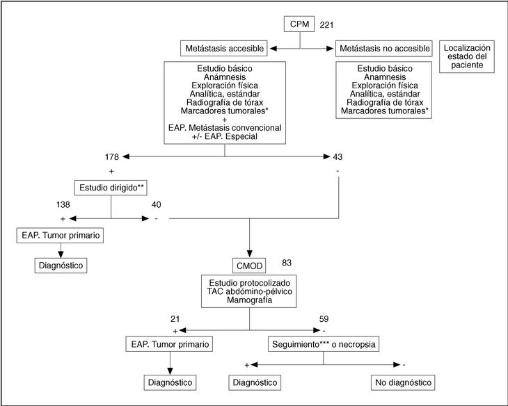
TABLA 1. Características de los pacientes, presentación metastásica y diagnóstico histológico
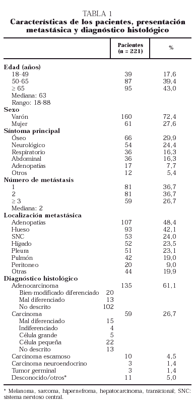
TABLA 2. Rendimiento del estudio básico en el cáncer de presentación metastásica
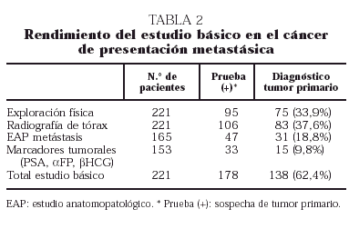
TABLA 3. Eficacia diagnóstica de los marcadores tumorales*
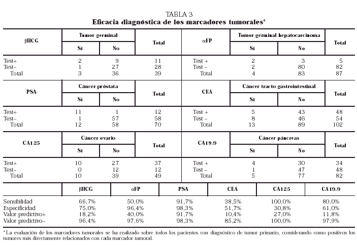
TABLA 4. Diagnóstico de tumor primario
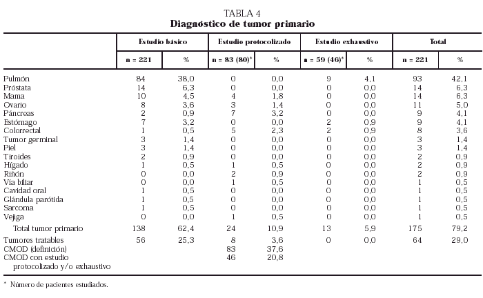
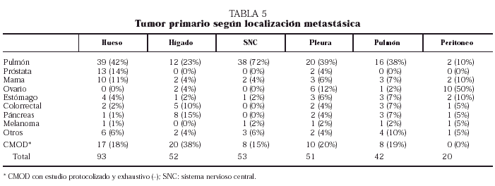
TABLA 5. Tumor primario según localización metastásica
Show moreShow less
Introducción. La aplicación de un algoritmo diagnóstico en el cáncer de presentación metastásica (CPM) podría facilitar, con un considerable ahorro de tiempo y exploraciones, llegar al diagnóstico de aquellos tumores primarios tratables. Material y métodos. Entre enero de 1992 y abril de 1997 se estudiaron de forma prospectiva todos los pacientes (pts) ingresados con el diagnóstico de CPM. Se les aplicó un estudio básico consistente en una historia clínica, un examen físico completo, una analítica estándar con marcadores tumorales y una radiografía de tórax. Se etiquetaron de cáncer metastásico de origen desconocido (CMOD) los pts con un estudio básico negativo, y en éstos se realizó un estudio protocolizado tomografía axial computarizada (TAC) (abdominopélvico y mamografía en mujeres). Aquellos pts en los que, tras la aplicación del estudio básico y el protocolizado no se detectó el tumor primario, fueron sometidos a un estudio exhaustivo a fin de validar la eficacia del algoritmo diagnóstico. Resultados. Se incluyeron 221 pts. La edad media era de 63 años (23-82). El síntoma principal fue óseo (30%), neurológico (24%), torácico (16%) y abdominal (16%). El estudio básico resultó positivo en 138 pts (62,4%); de éstos, la radiografía de tórax y la exploración física aportaron el mayor número de diagnósticos. La histología de las metástasis contribuyó al diagnóstico definitivo en 31 pts. Sólo el antígeno prostático específico (PSA) presentó una alta sensibilidad y especificidad. Fueron etiquetados de CMOD 83 pts. El estudio protocolizado diagnosticó el tumor primario en 24 pts (30%), 20 por TAC abdominal y 4 por mamografía; de éstos, 8 pts se consideraron tratables. En los 59 pts restantes se aplicó un estudio exhaustivo, hallándose el diagnóstico en 13; sin embargo, ninguno se consideró claramente merecedor de un tratamiento específico. Finalmente 47 pts (21%) quedaron sin diagnóstico. Los tumores primarios predominantes fueron pulmón (42%), próstata (6%) y mama (6%). Las localizaciones metastásicas más frecuentes fueron hueso (42%), sistema nervioso central e hígado (24%), y la histología, adenocarcinoma (61%) y carcinoma indiferenciado (15%). Conclusiones. El cáncer de pulmón y el CMOD representaron el 62% de los CPM. El estudio básico puede orientar dos tercios de los casos, siendo la exploración física y la radiografía de tórax las que tienen mayor rentabilidad diagnóstica. La histología de las metástasis y el PSA son de capital importancia. Un estudio protocolizado basado en la TAC abdominopélvica y la mamografía en mujeres puede identificar el resto de tumores tratables.
Palabras clave:
carcinoma metastásico de origen desconocido, algoritmo diagnóstico
Background. The use of a diagnostic algorithm for metastatic cancer presentation (MCP) might enhance the diagnosis of primary tumors amenable to treatment with considerable savings both in time and diagnostic examinations. Materials and methods. From January 1992 to April 1997, all patients admitted with the diagnosis of MCP were prospectively studied. From each patient, a basic study consisting in a clinical interview, complete physical examination, standard blood testing with tumoral markers and chest X-ray were obtained. Patients with a negative basic study were classified as having a metastatic cancer of unknown origin (MUO); in these patients, a protocolized study (abdominal CT scan and mammography among women) were performed. Patients who after the application of the basic and protocolized studies had no primary tumor detected underwent an exhaustive investigation in order to validate the efficiency of the diagnostic algorithm. Results. Two hundred twenty-one patients were included in the study. The mean age of patients was 63 years (range: 23-82). The main symptom was of bone (30%), neurological (24%), thoracic (16%) and abdominal (16%) origin. The basic study was positive for 138 patients (62.4%), with chest X-ray and physical examination yielding the highest number of diagnoses among these patients. The histology of metastases contributed to the definite diagnosis in 31 patients. Only PSA had a high sensitivity and specificity. Eighty-three patients were classified as MUO. The protocolized study diagnosed the primary tumor in 24 patients (30%), 20 by abdominal CT scan and four by mammography; eight of these patients were deemed to be amenable to treatment. The remaining 59 patients underwent an exhaustive study, and a diagnosis was made in 13; nevertheless, none of them was considered candidate for a specific treatment. Finally, 47 patients (21%) remained undiagnosed. The predominant primary tumors included sites at the lung (42%), prostate (6%) and breast (6%). The most common metastatic locations included bone (42%), central nervous system and liver (24%), and the most common histological types were adenocarcinoma (61%) and undifferentiated carcinoma (15%). Conclusions. Lung cancer and MUO represented 62% of MCP. The basic study oriented in two thirds of cases, and the physical examination and chest X-ray showed the highest diagnostic yield. The histology of metastases and PSA had a key, diagnostic relevance. A protocolized study based on abdominal CT scan and mammography (females) can identify the remaining treatable tumors.
Keywords:
metastatic carcinoma of unknown origin, diagnostic algorithm
Article
These are the options to access the full texts of the publication Revista Clínica Española (English Edition)
Member
Si es usted socio de FESEMI siga los siguientes pasos:
Diríjase desde aquí a la web de la >>>FESEMI<<< e inicie sesión mediante el formulario que se encuentra en la barra superior, pulsando sobre el candado.

Una vez autentificado, en la misma web de FESEMI, en el menú superior, elija la opción deseada.

>>>FESEMI<<<
Purchase

